
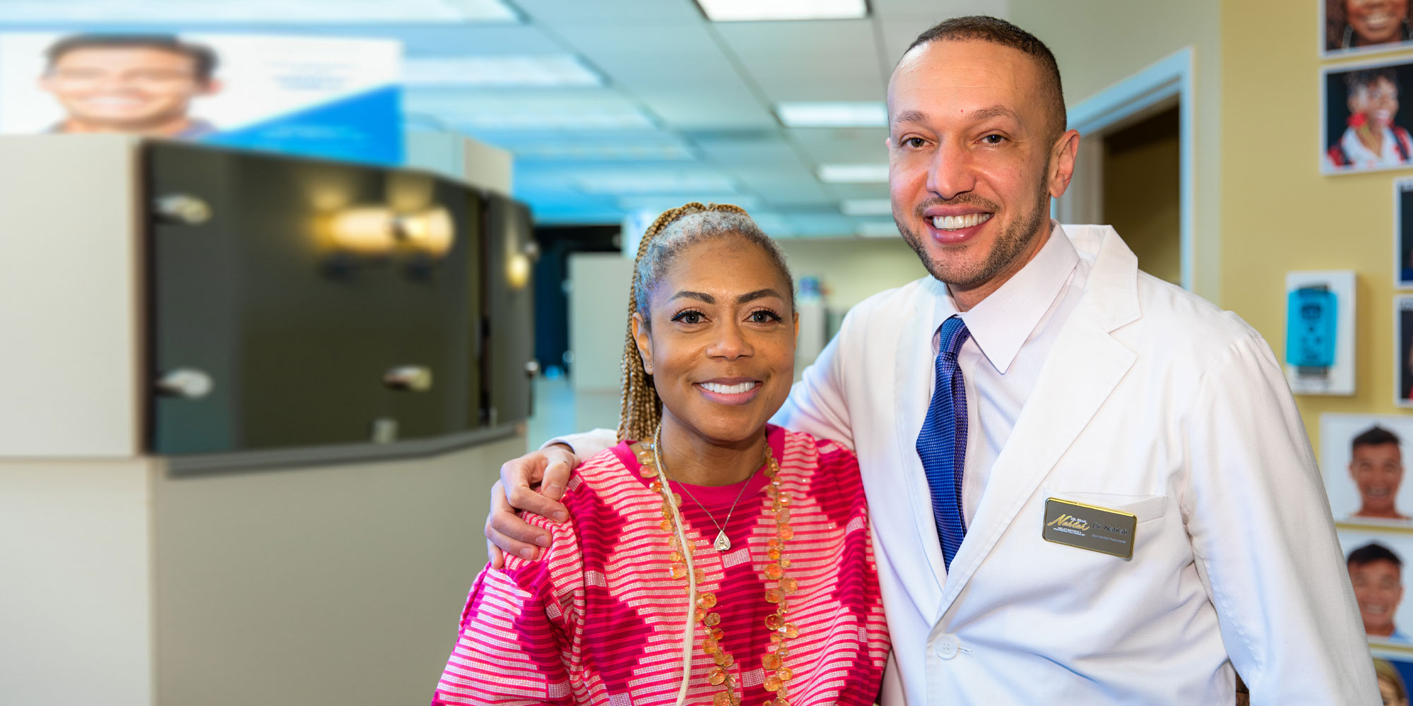
54 cases. No stock photography.
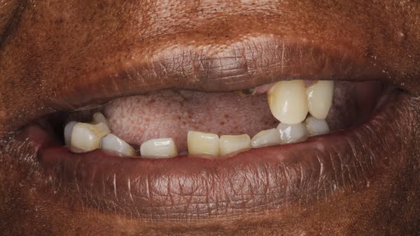
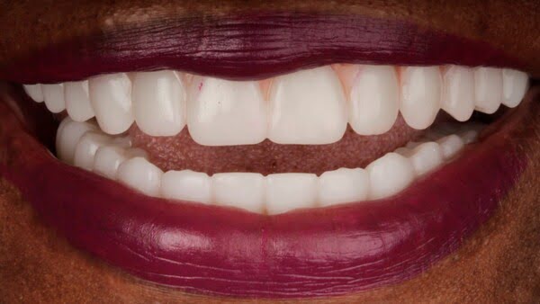
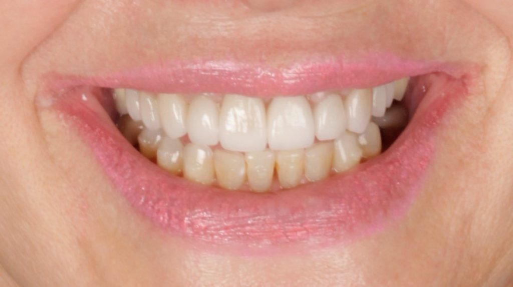
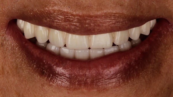
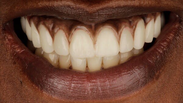
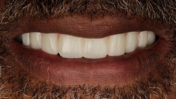
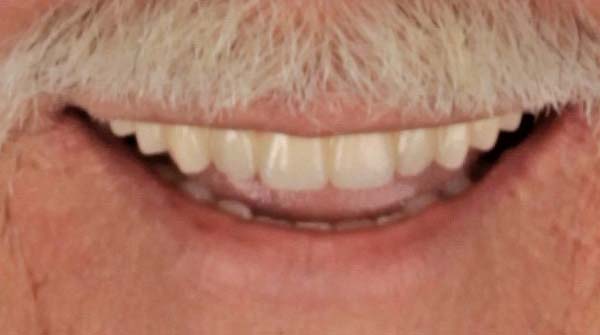
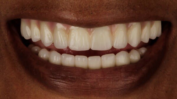
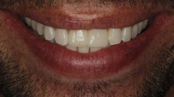
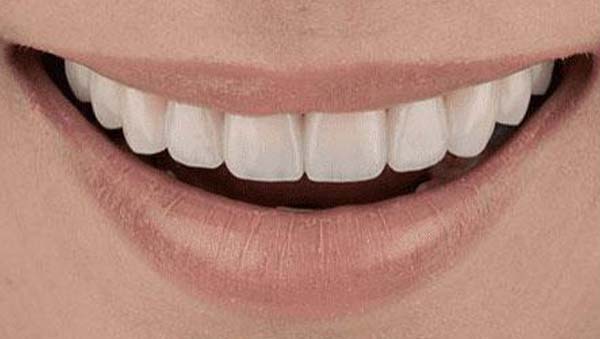
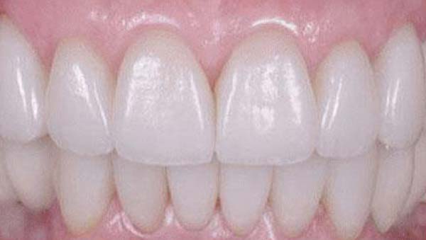
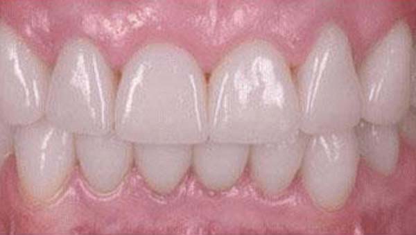
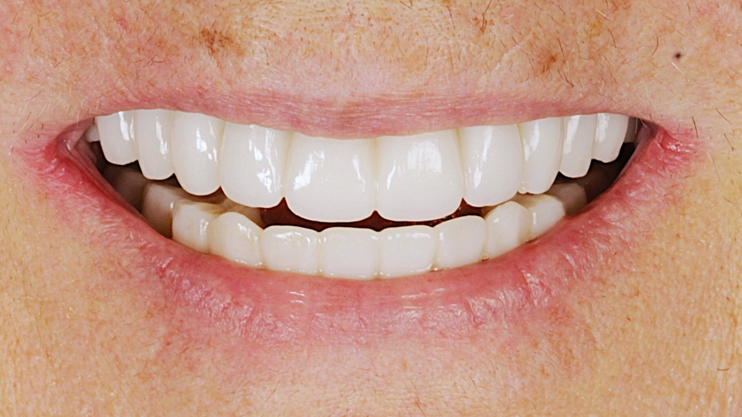
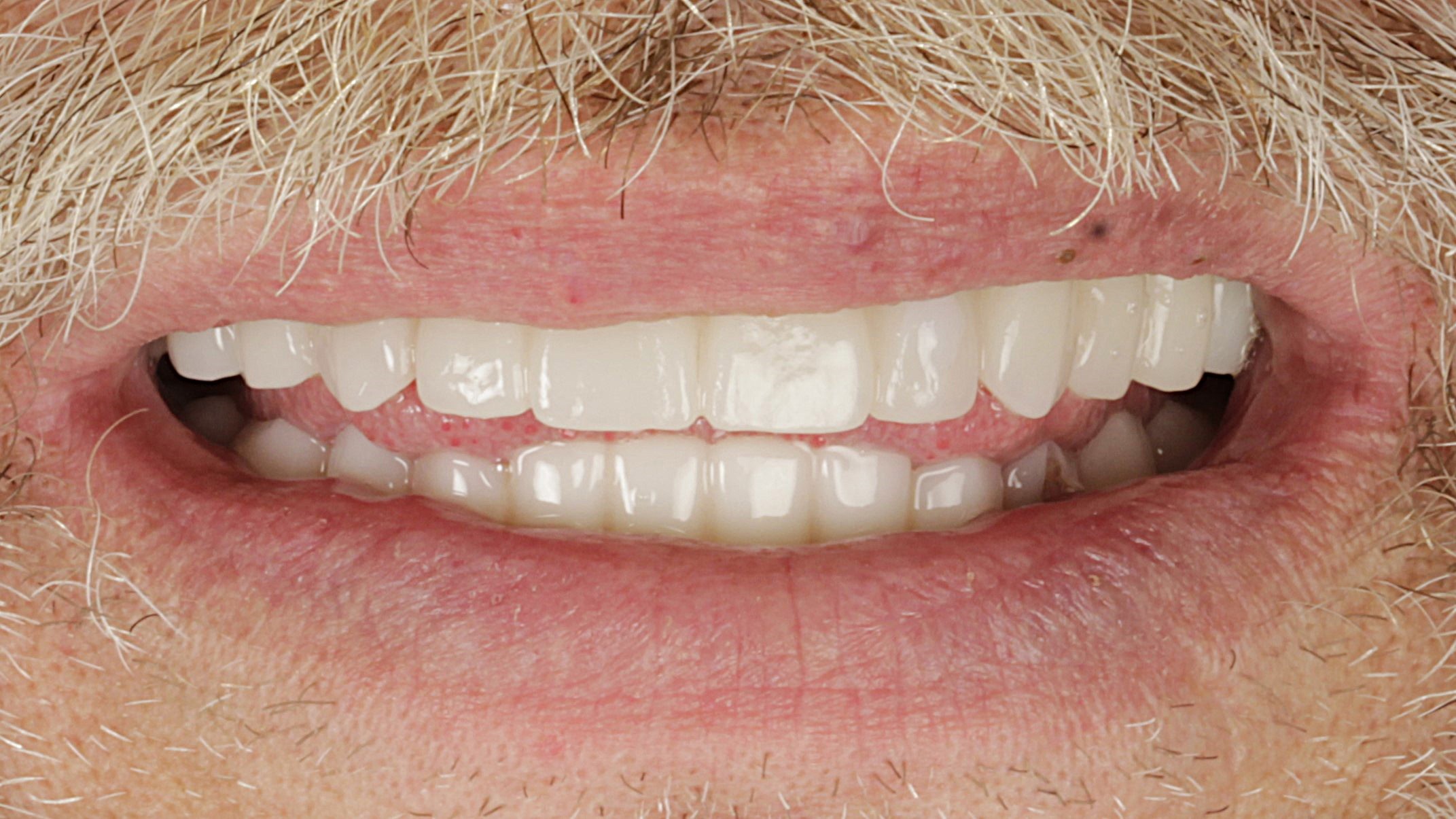
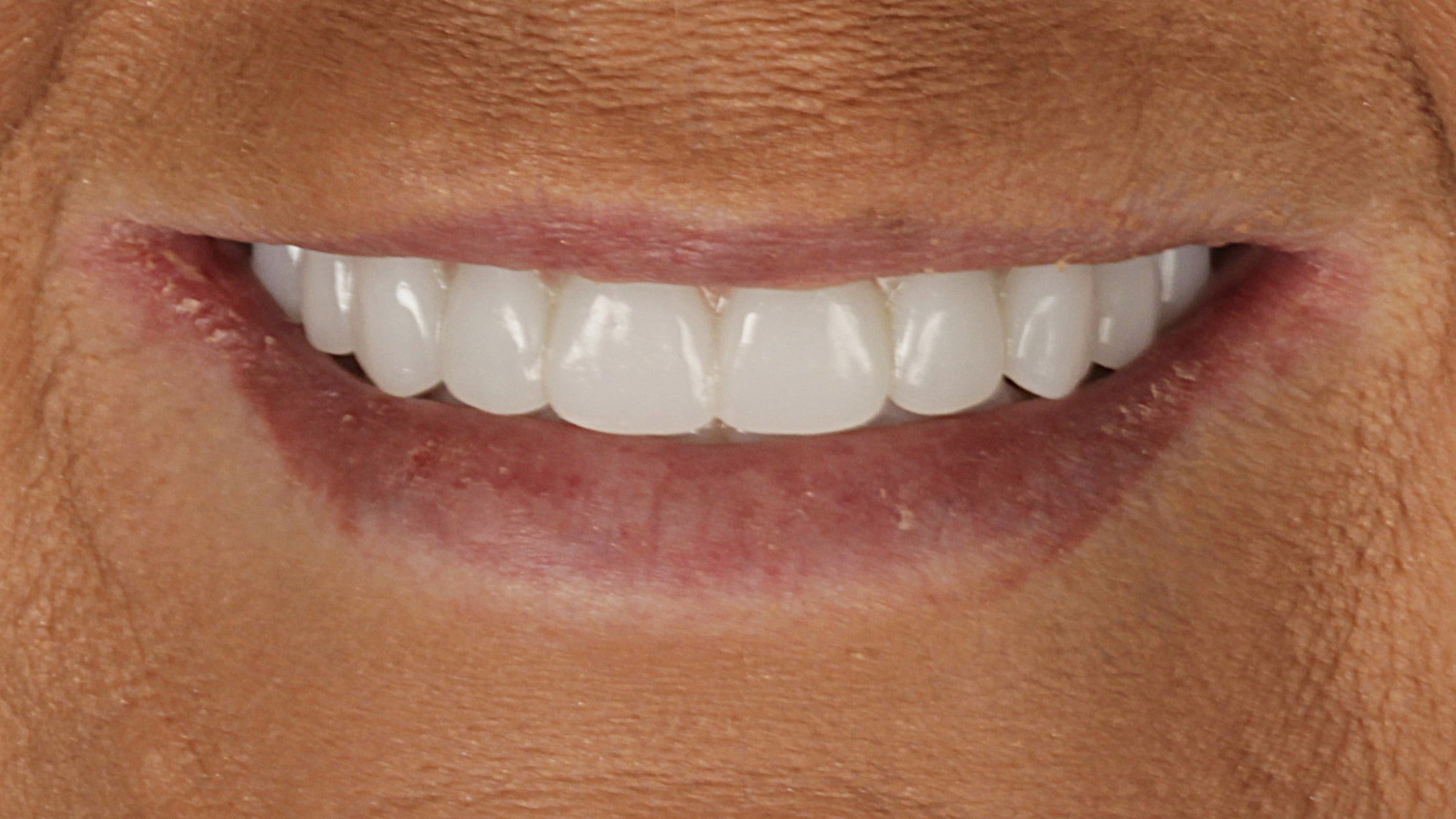
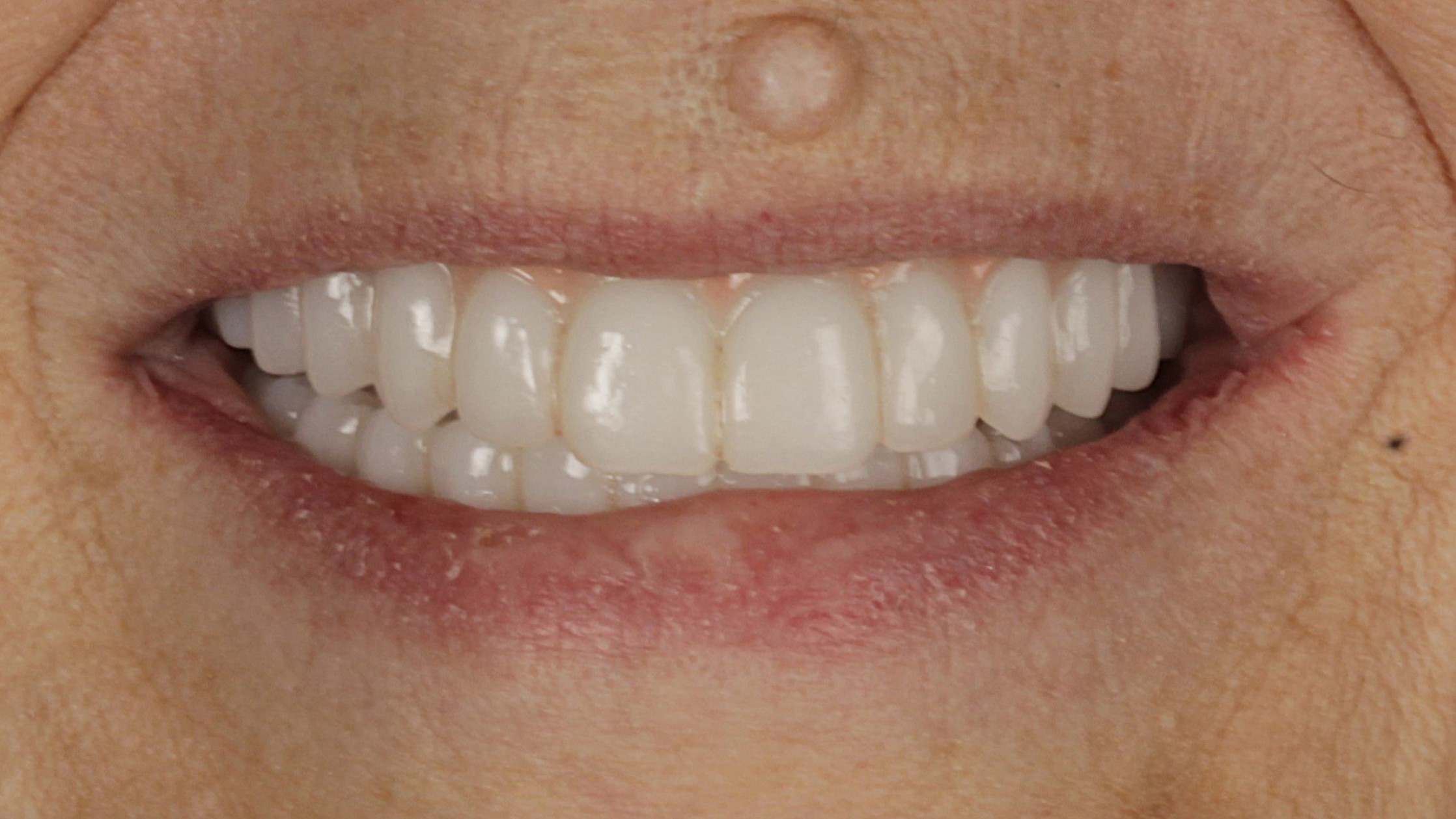
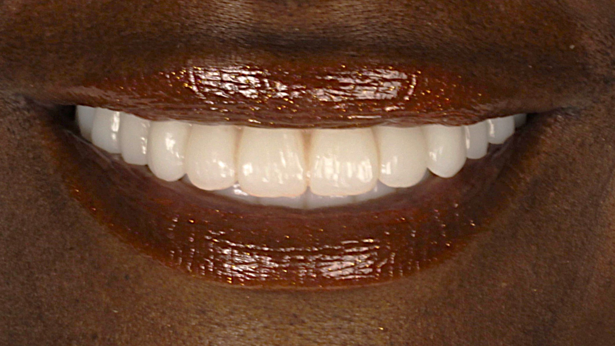
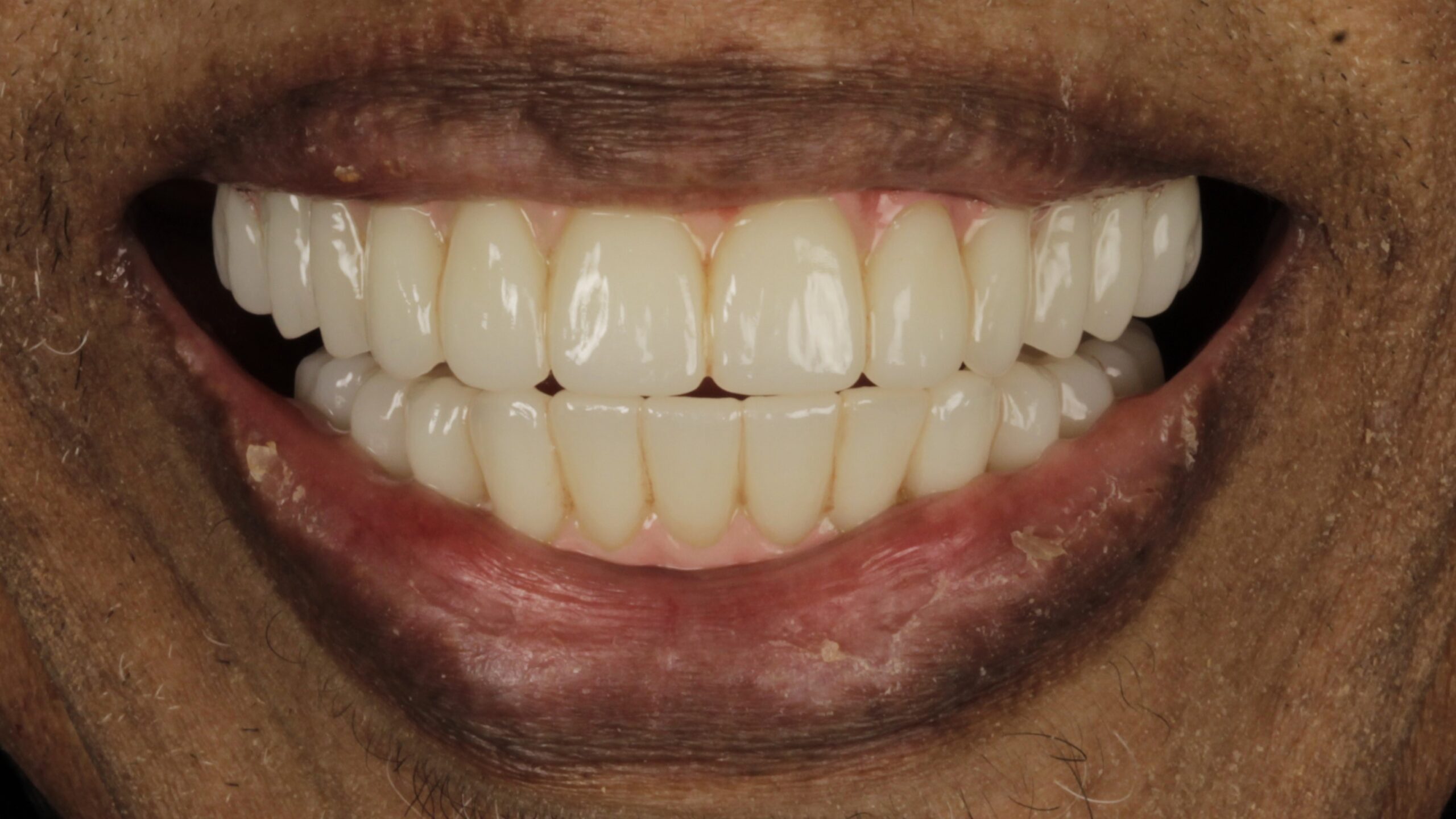
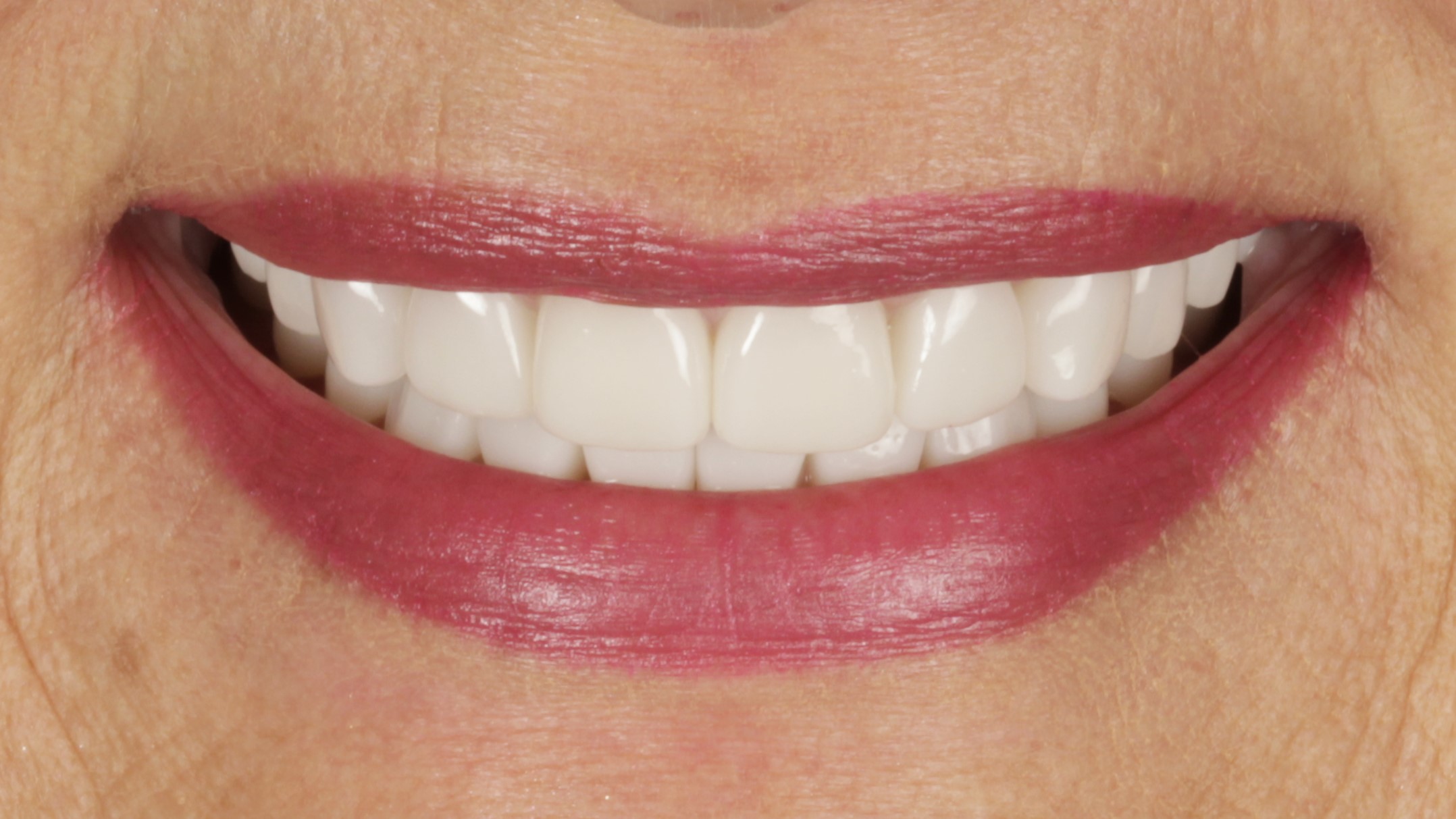
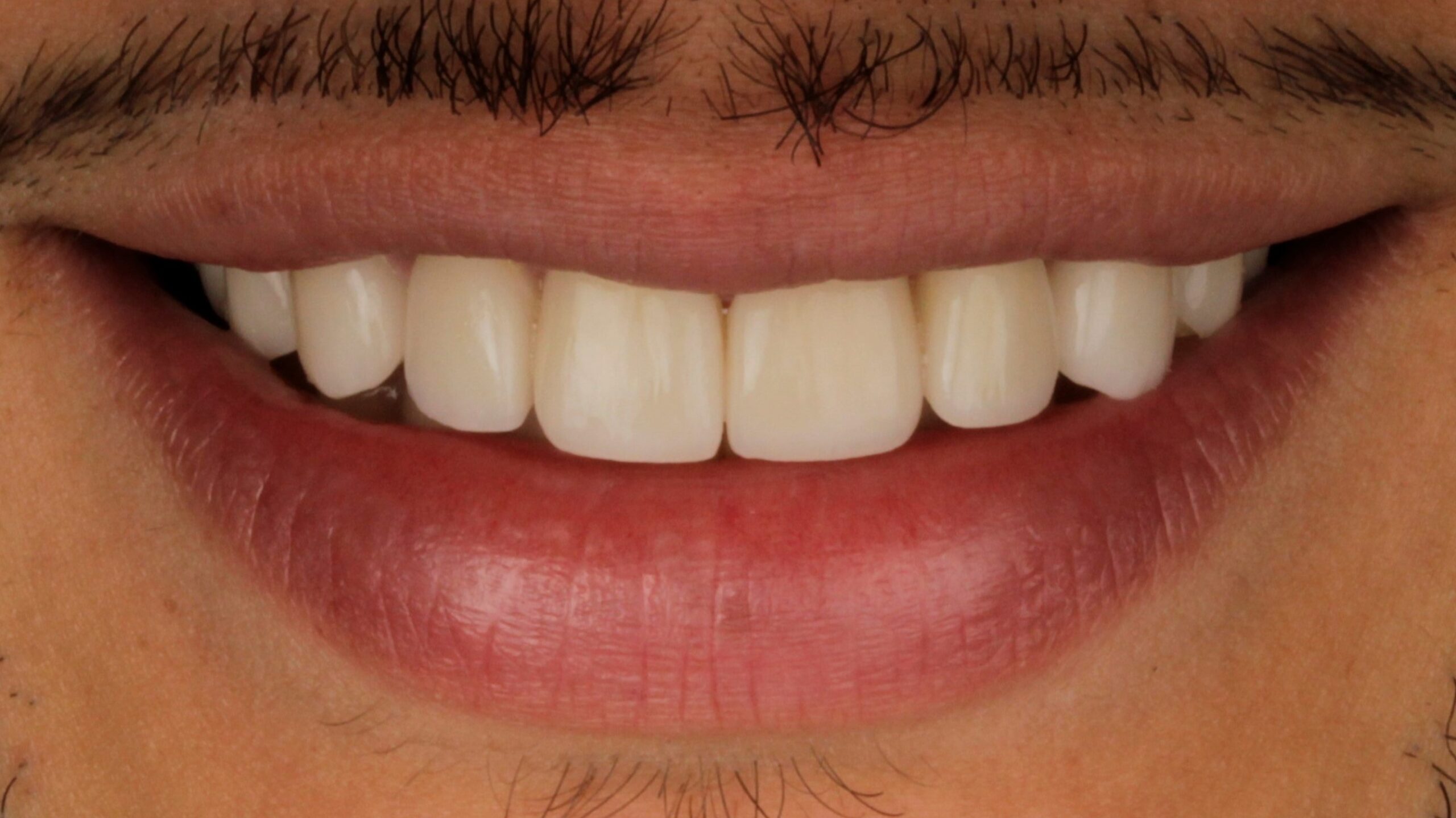
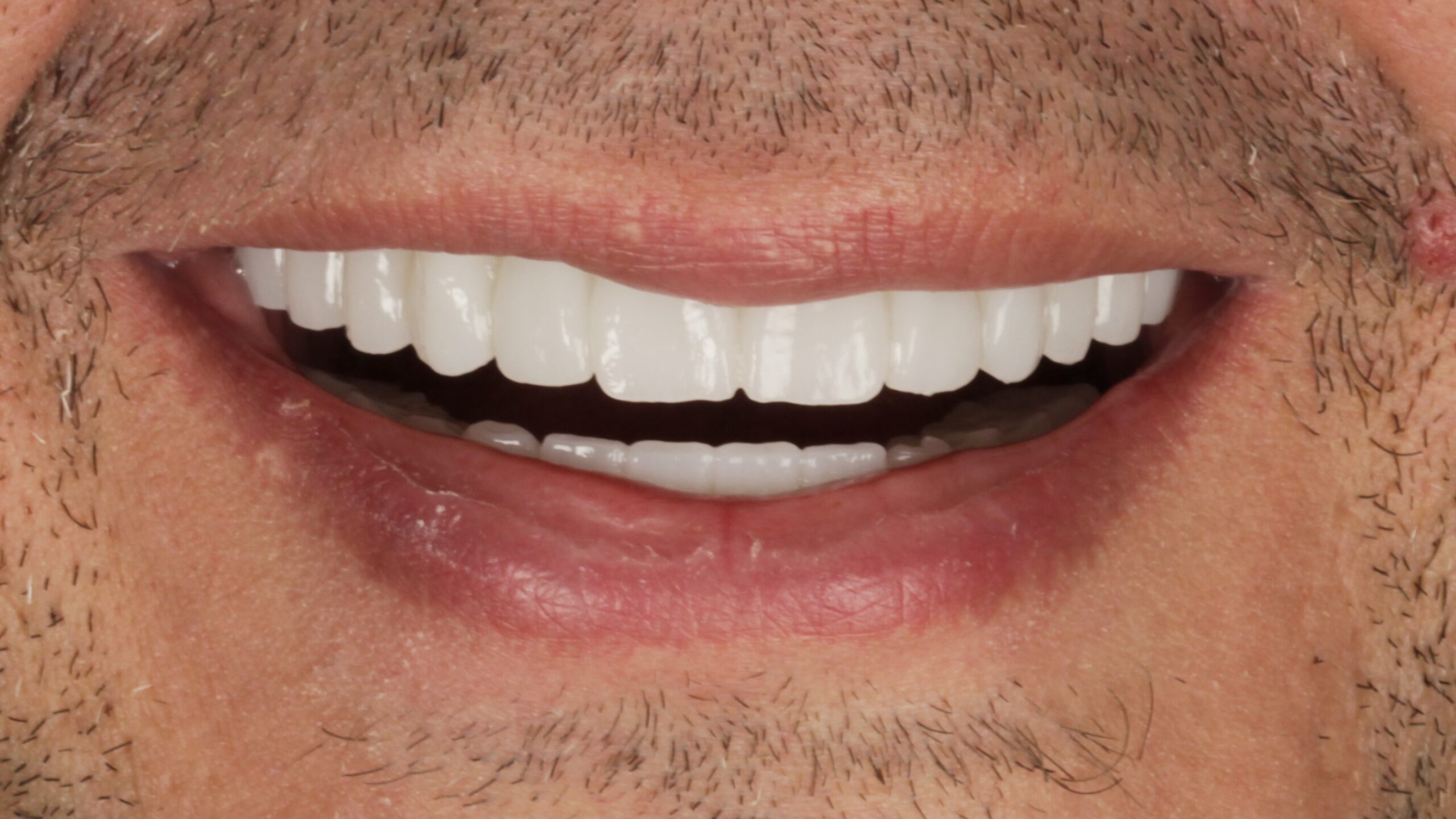
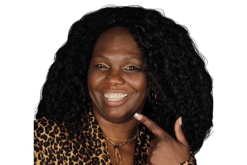
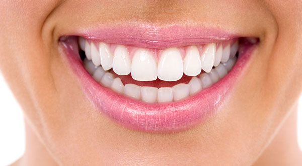
Every pair below was treated at this practice. Drag the line across any of them, or use the keyboard arrows once a case has focus.
Sorted by what was actually done.
 BeforeAfter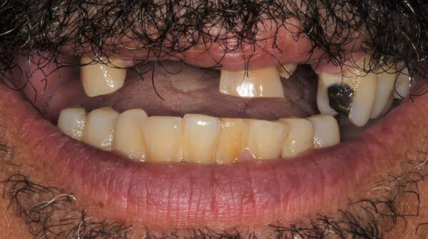
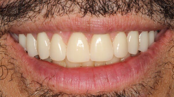 BeforeAfter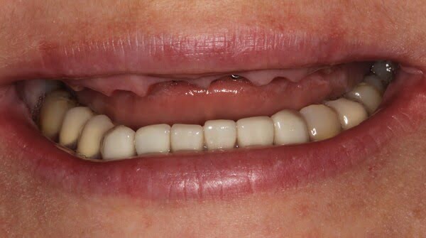
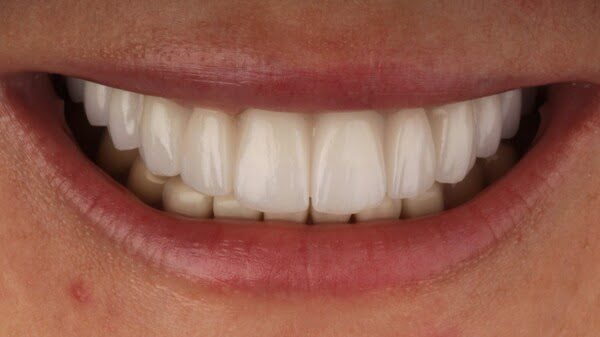 BeforeAfter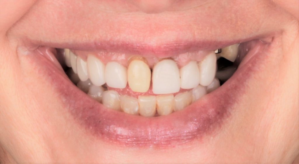
 BeforeAfter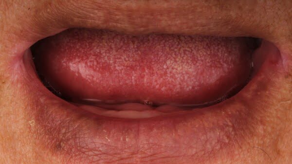
 BeforeAfter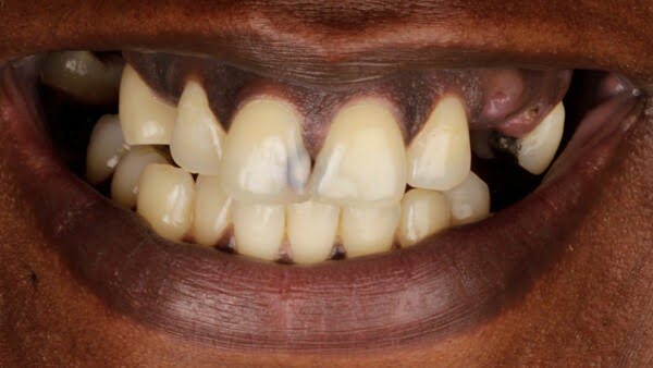
 BeforeAfter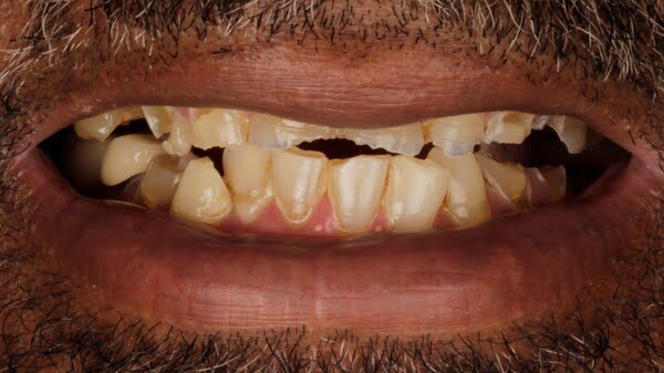
 BeforeAfter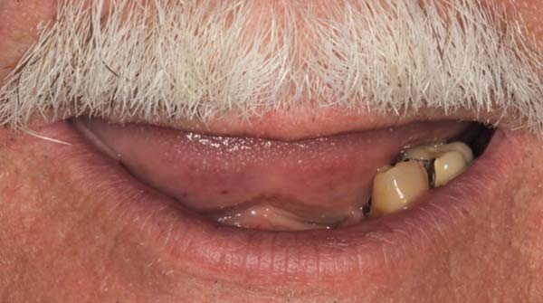
 BeforeAfter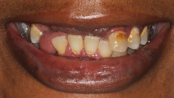
 BeforeAfter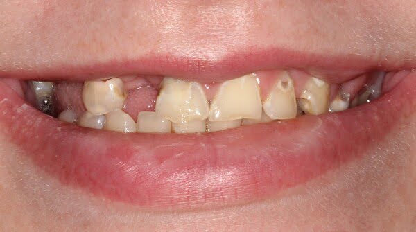
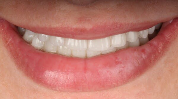 BeforeAfter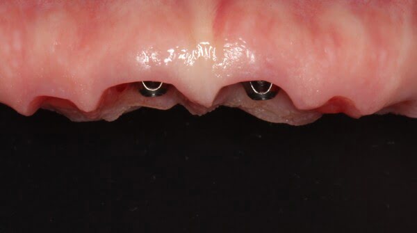
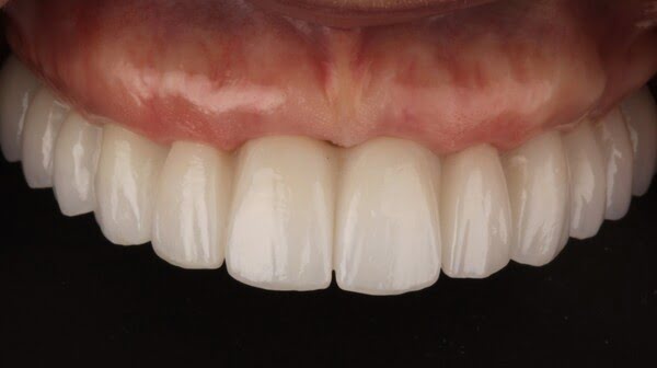 BeforeAfter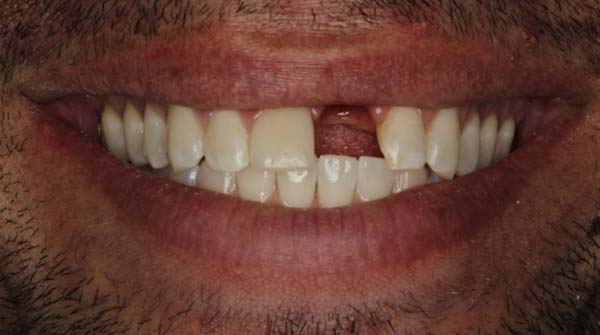
 BeforeAfter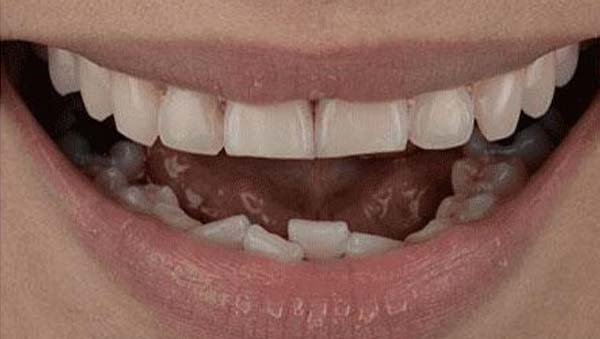
 BeforeAfter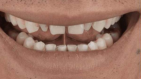
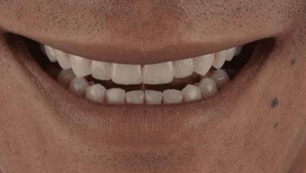 BeforeAfter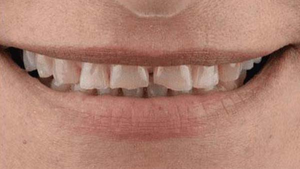
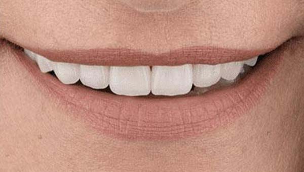 BeforeAfter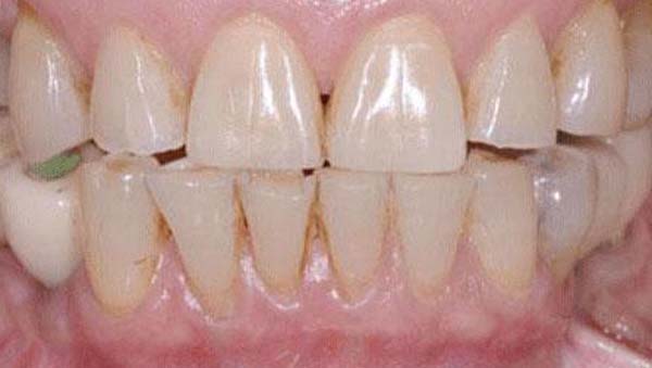
 BeforeAfter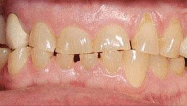
 BeforeAfter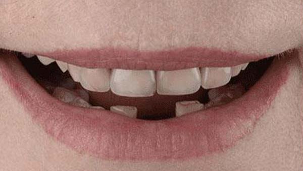
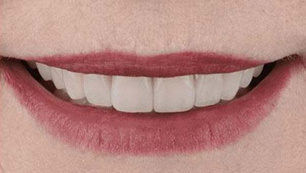 BeforeAfter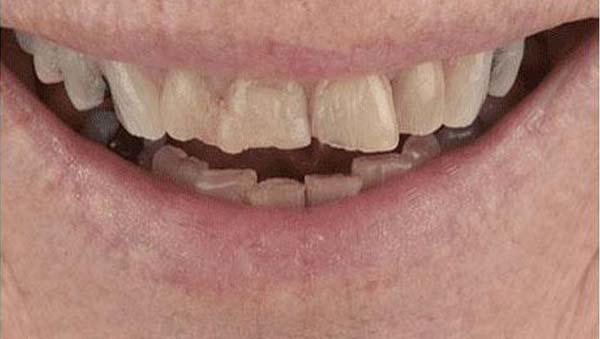
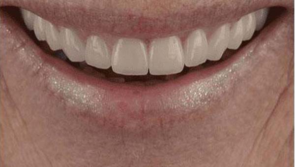 BeforeAfter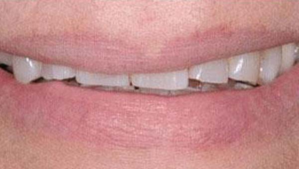
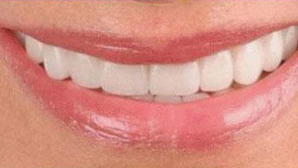 BeforeAfter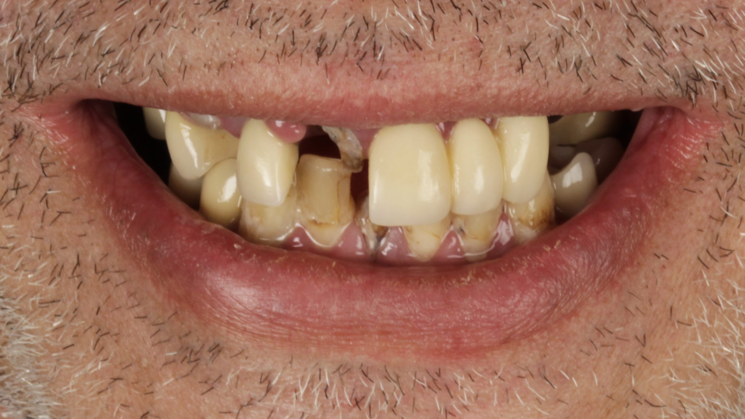
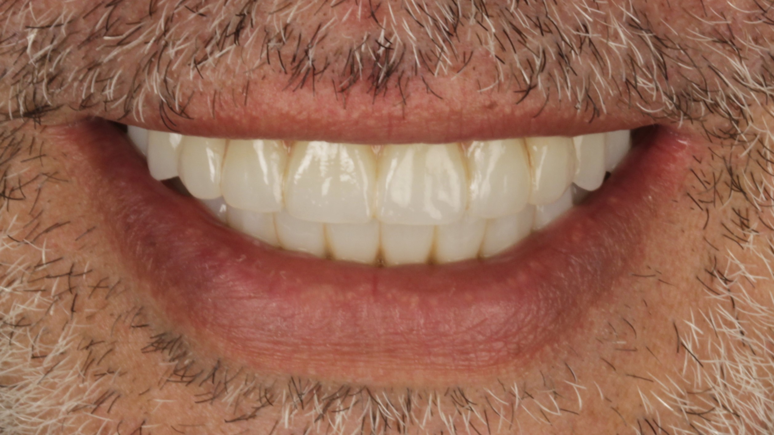 BeforeAfter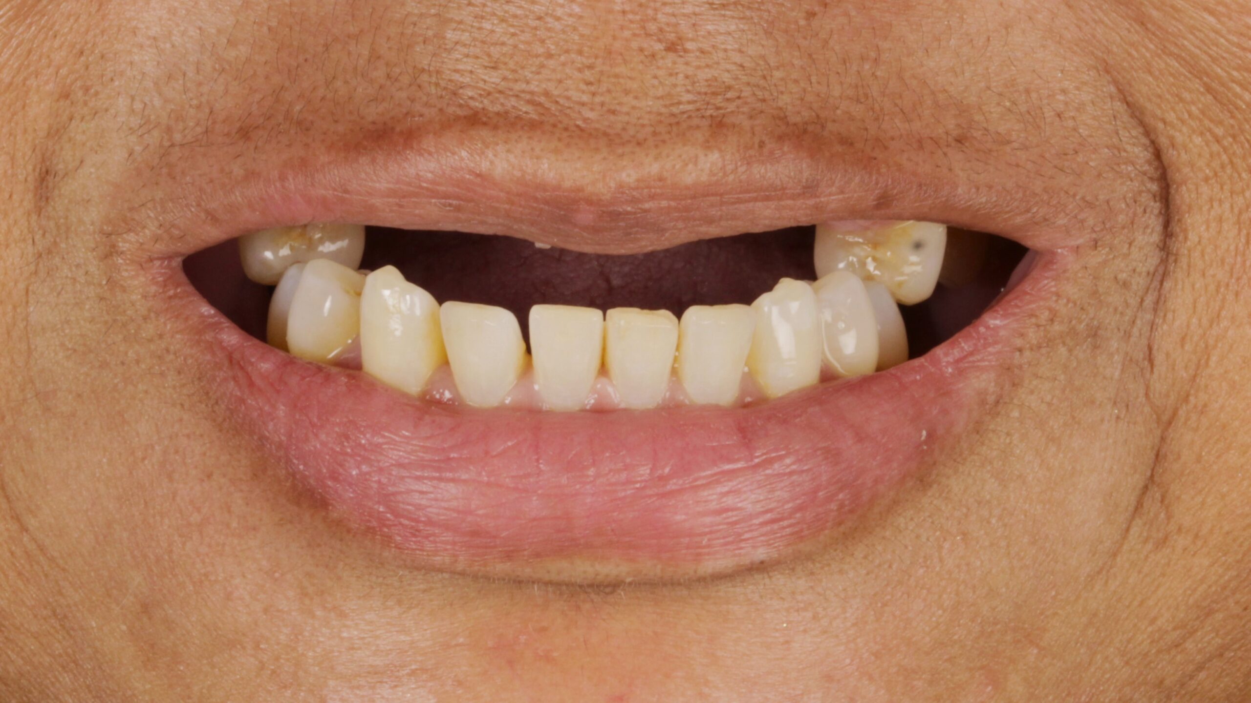
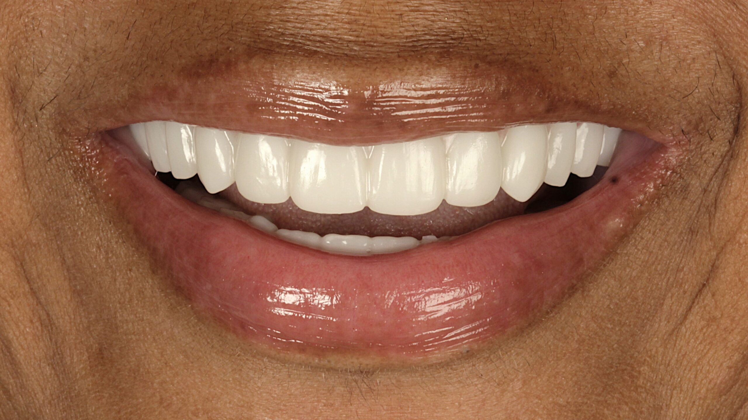 BeforeAfter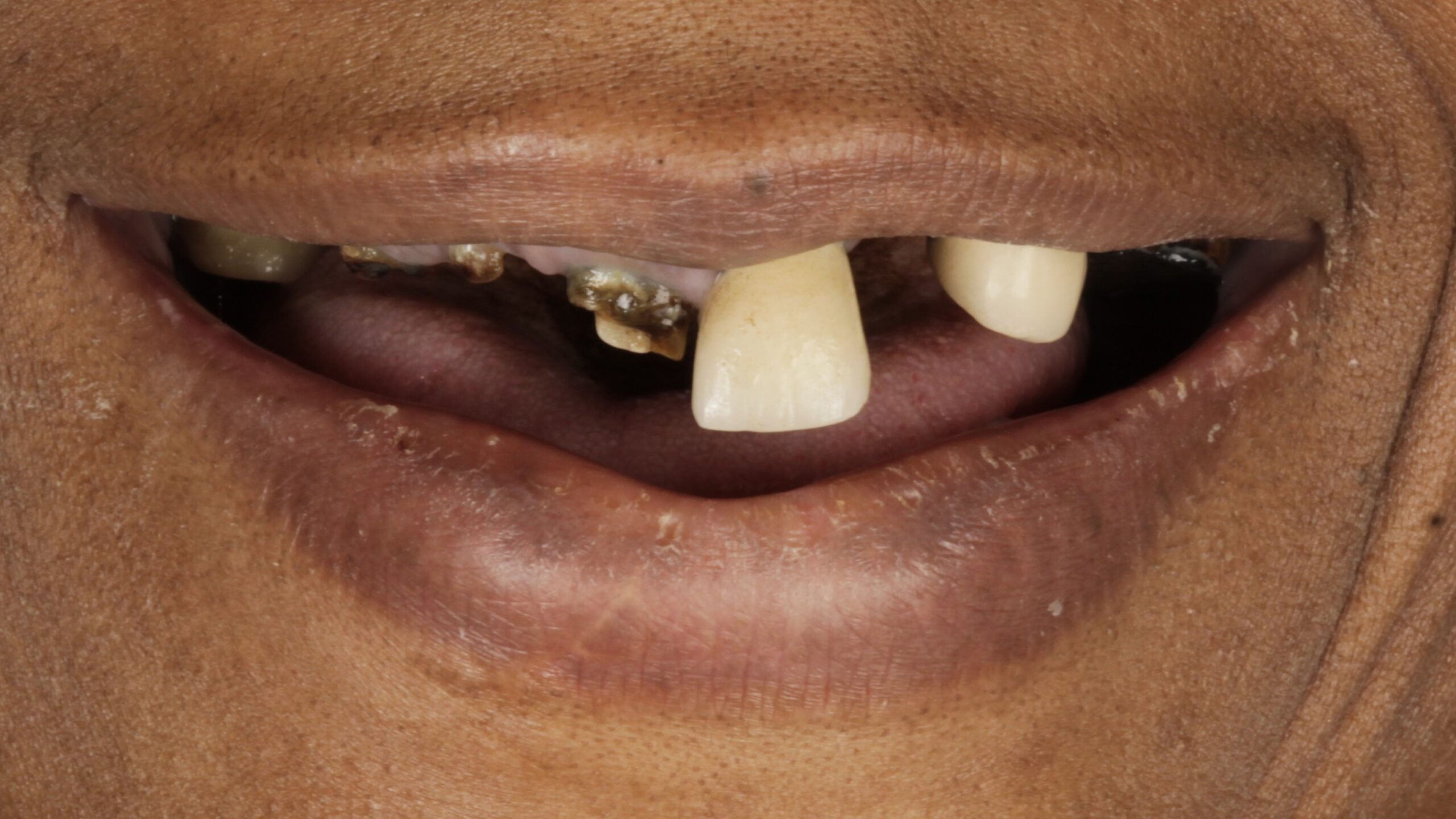
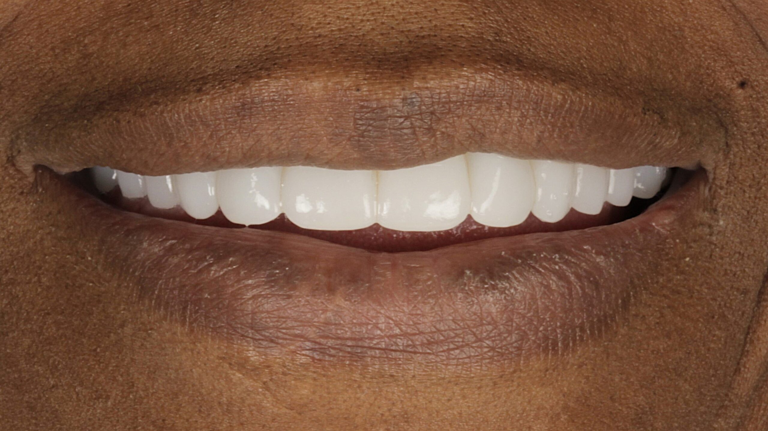 BeforeAfter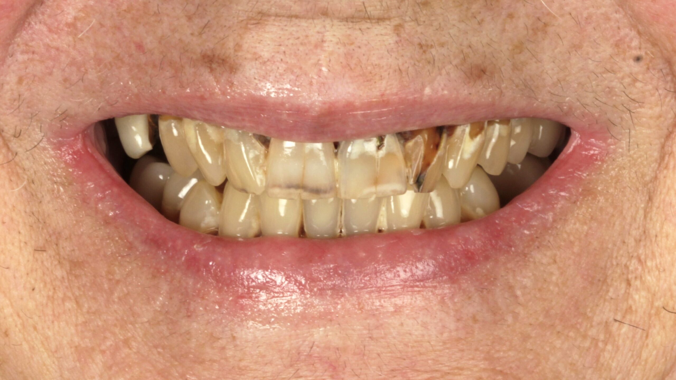
 BeforeAfter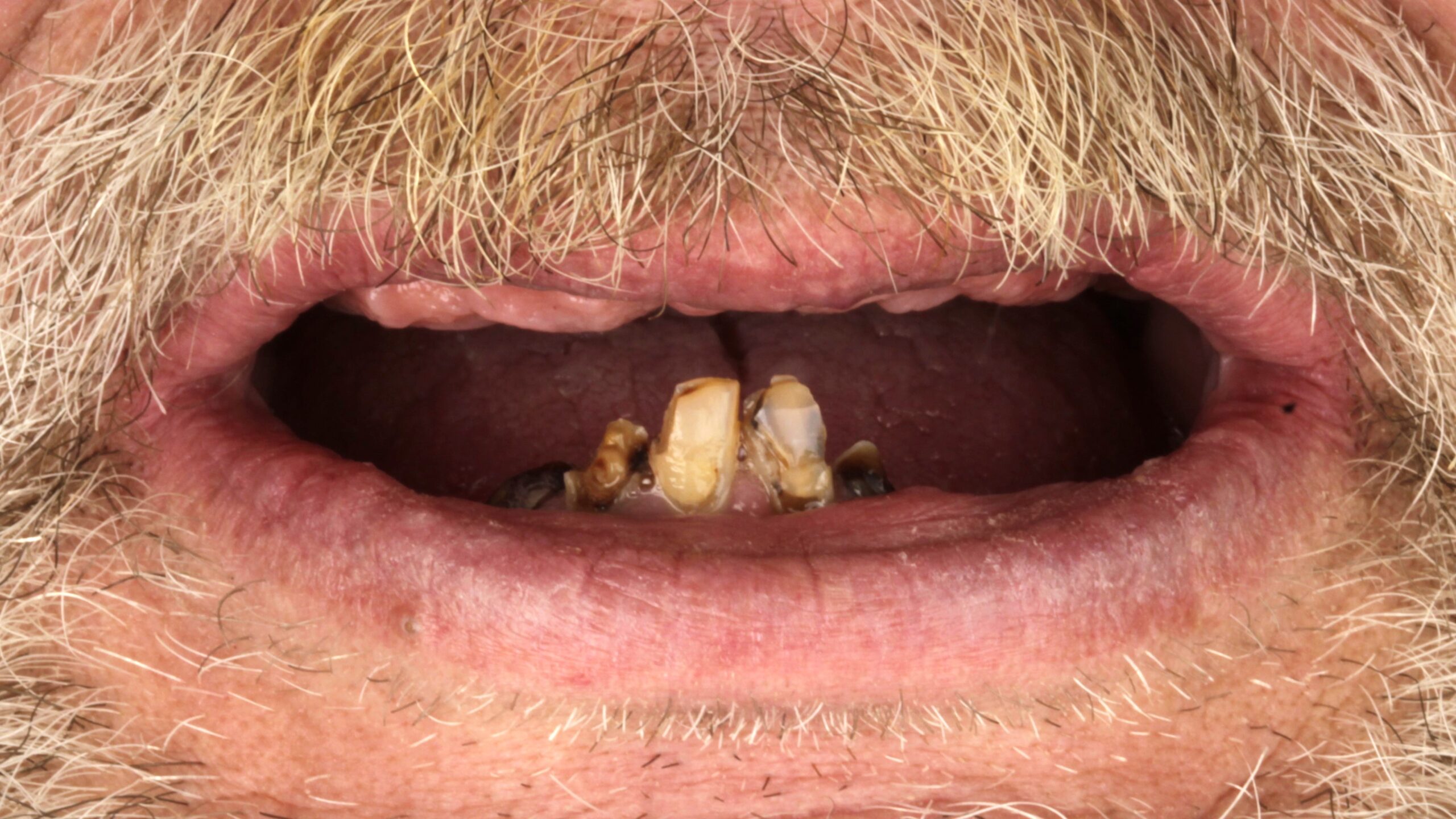
 BeforeAfter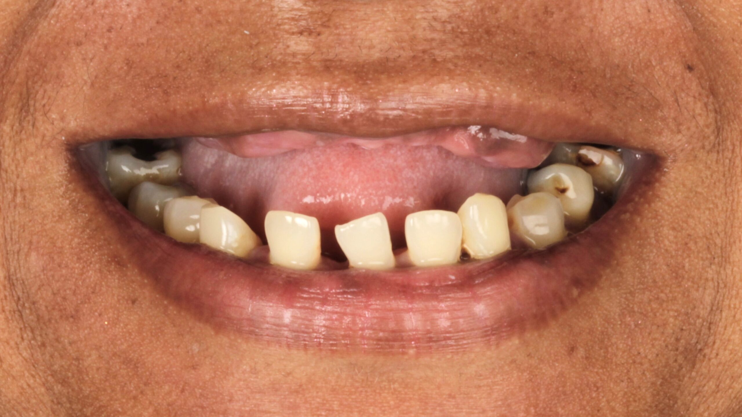
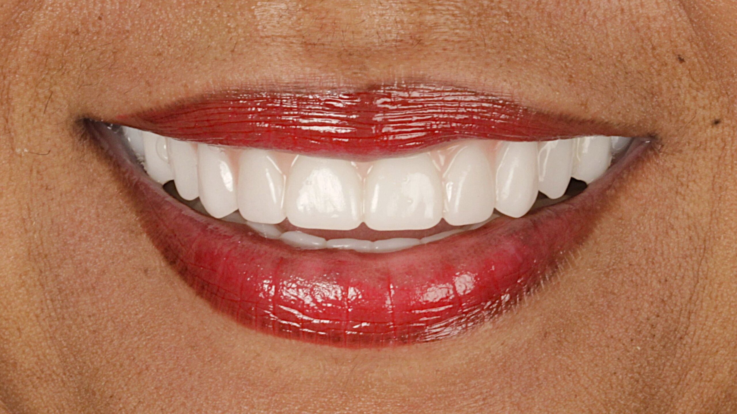 BeforeAfter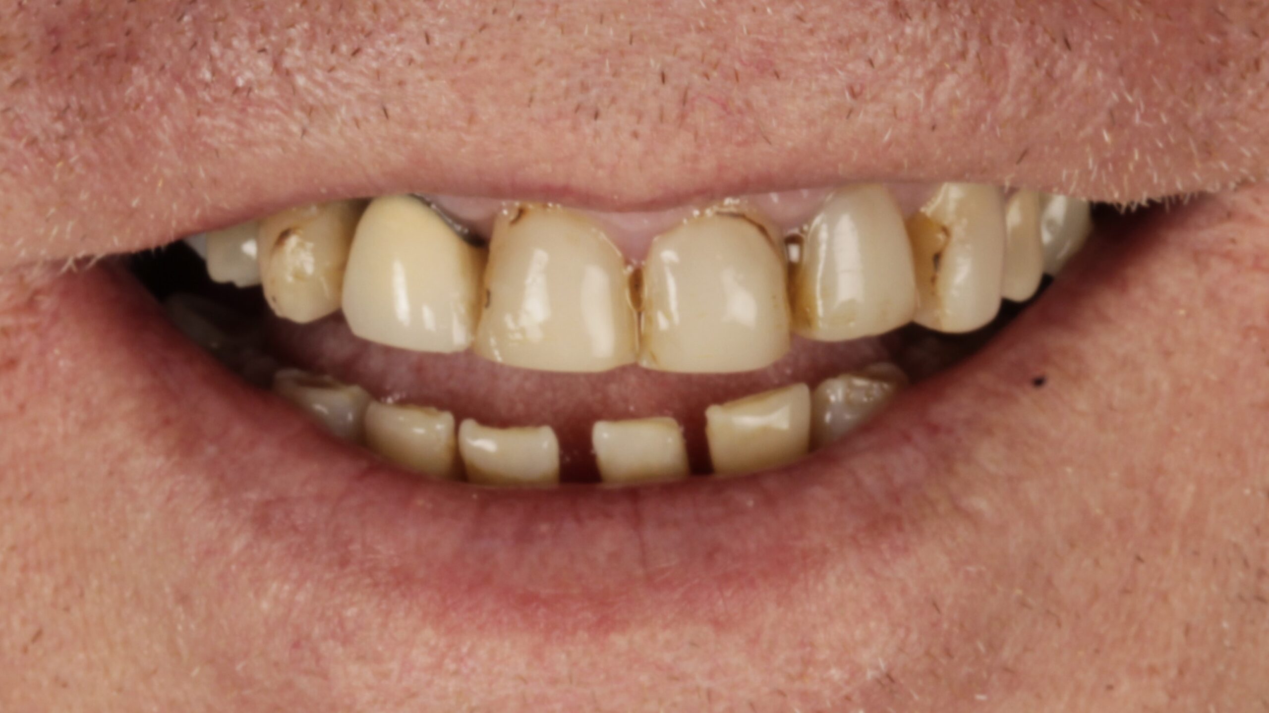
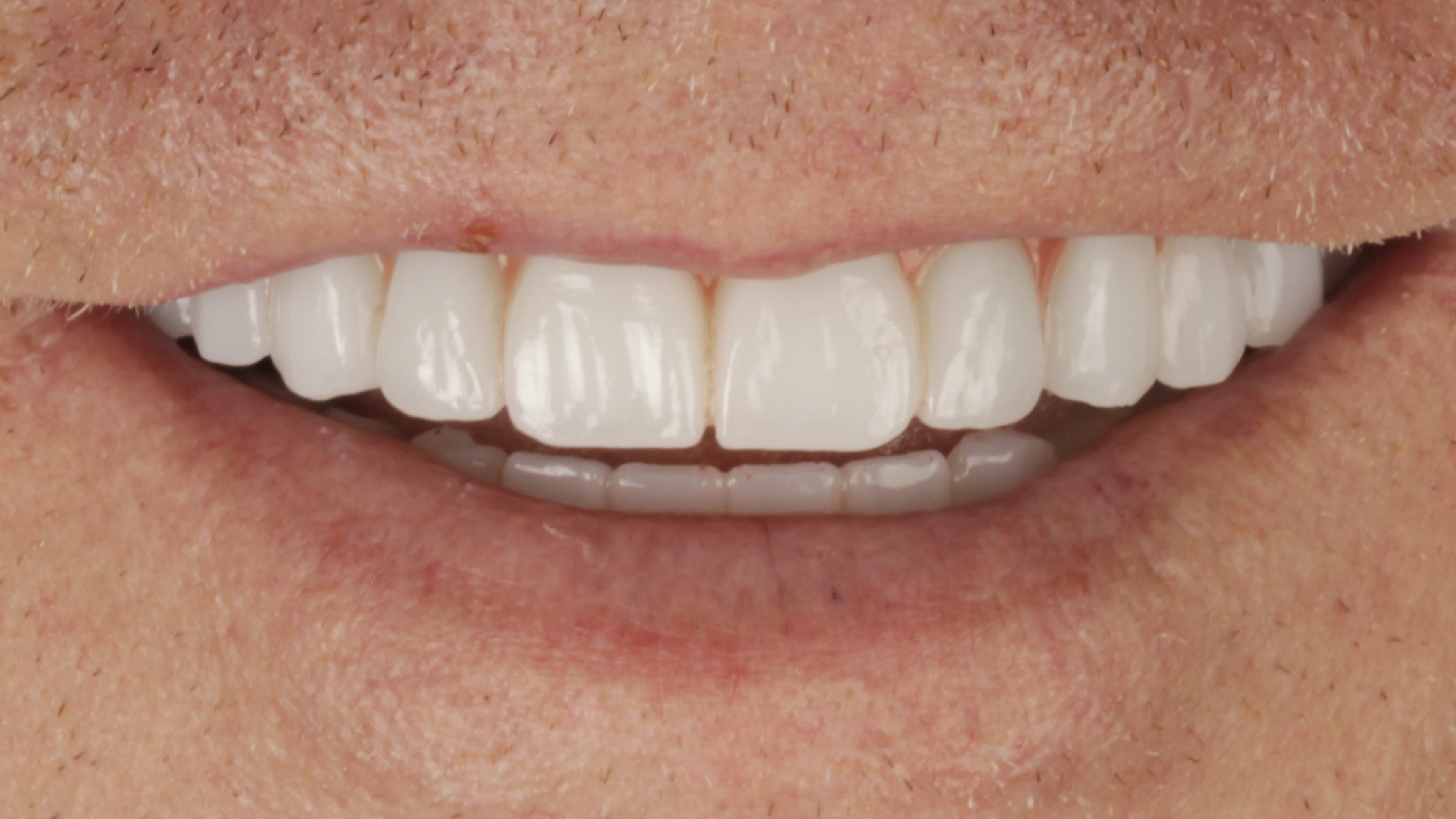 BeforeAfter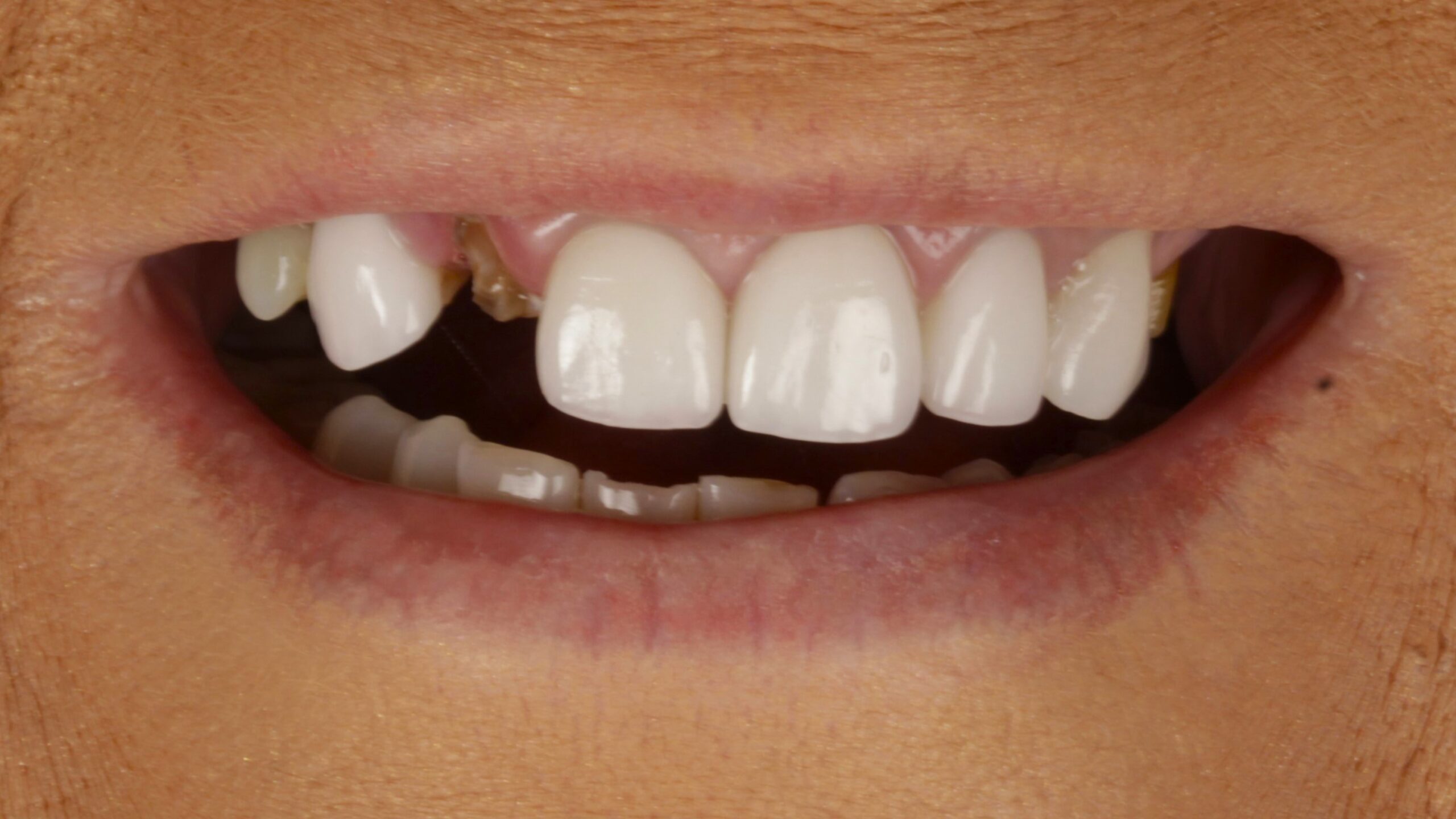
 BeforeAfter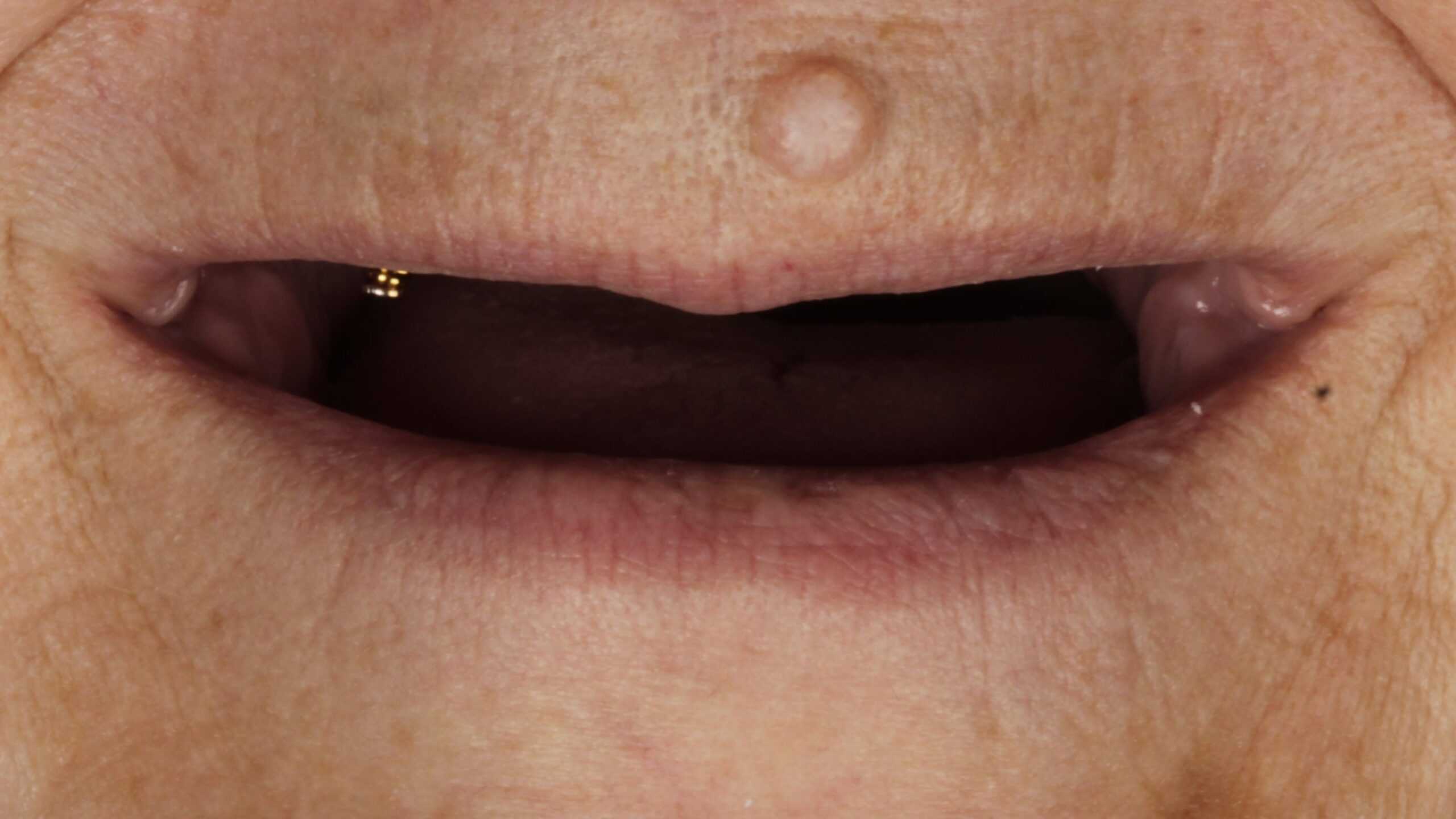
 BeforeAfter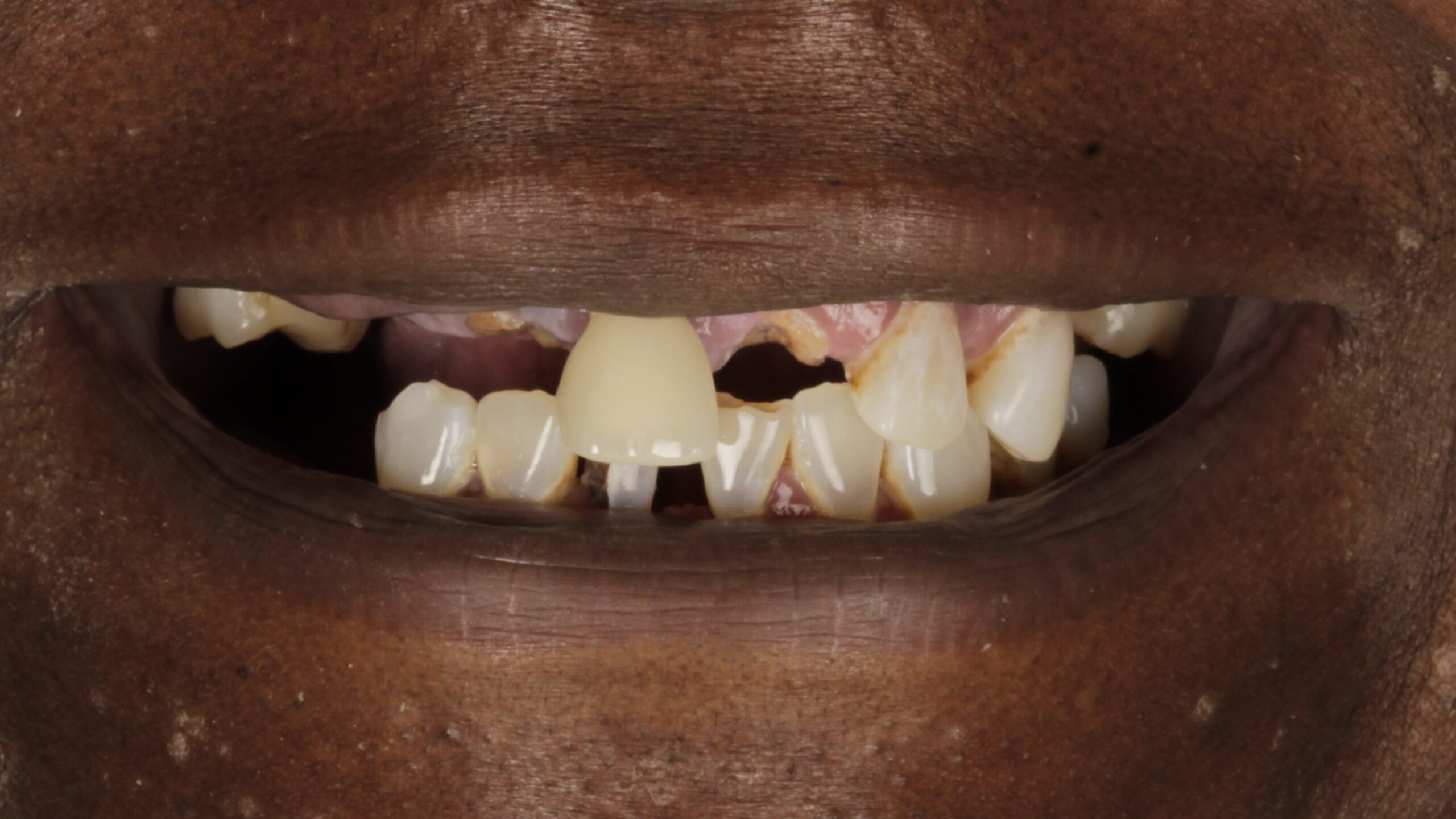
 BeforeAfter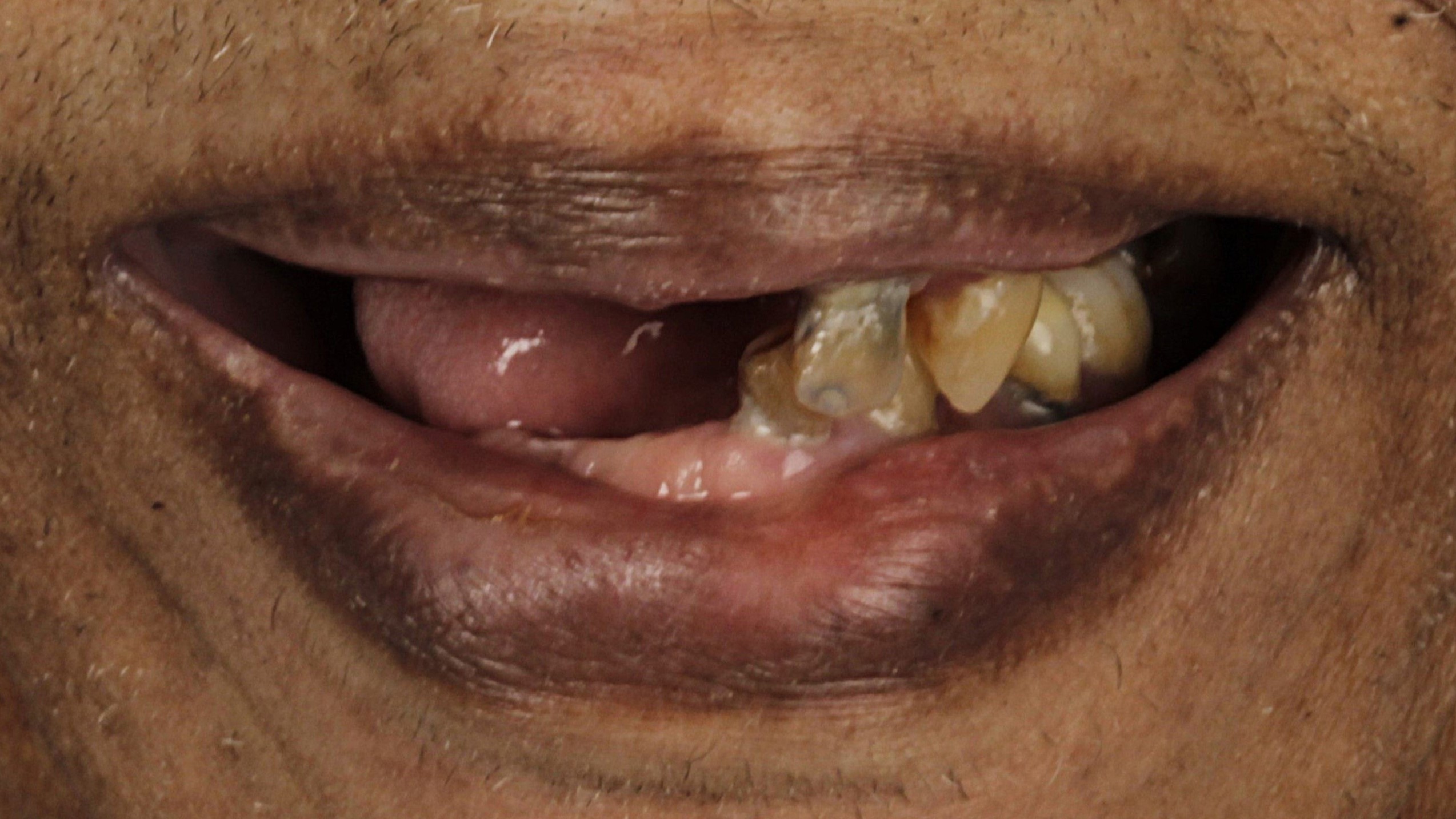
 BeforeAfter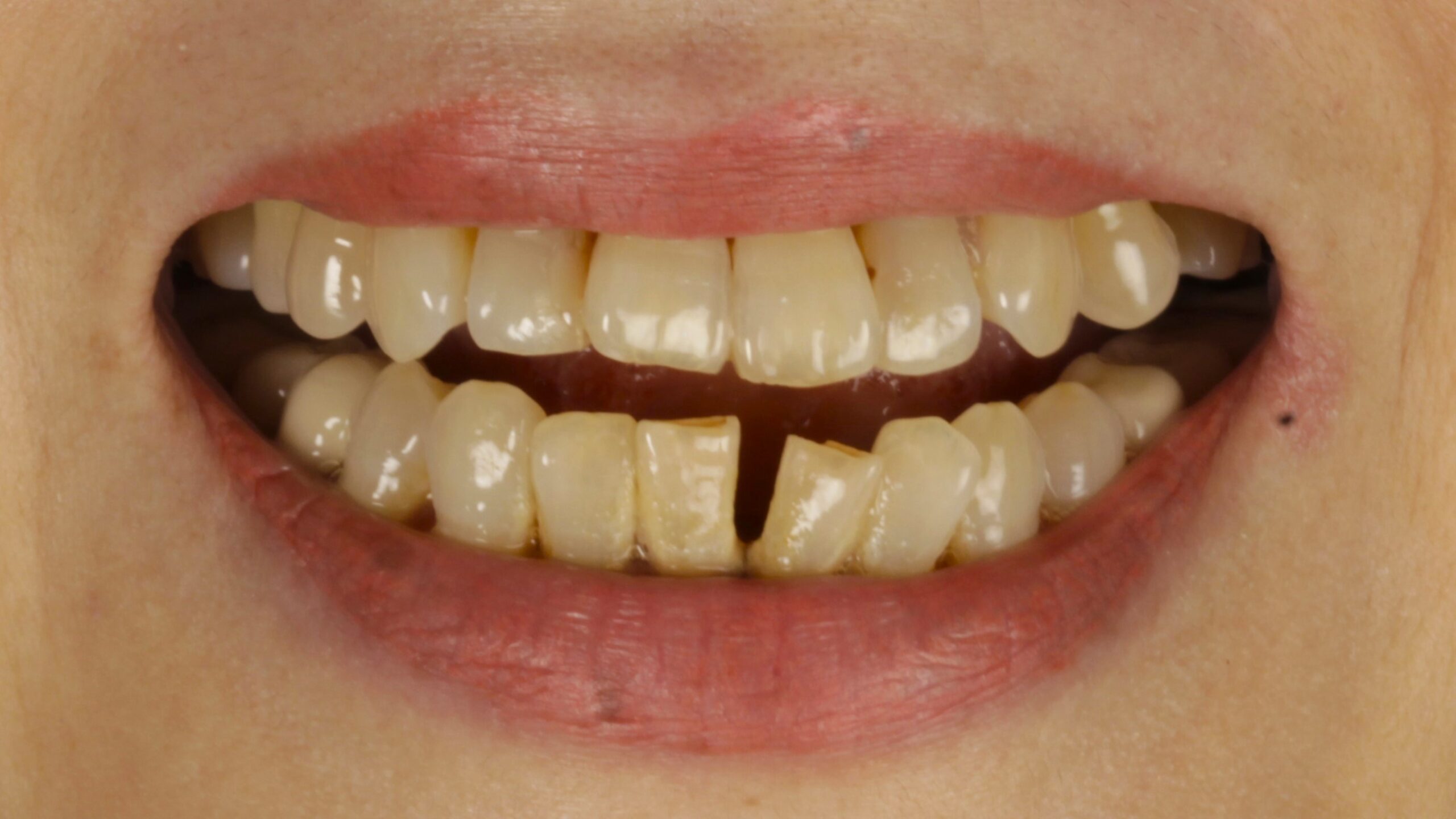
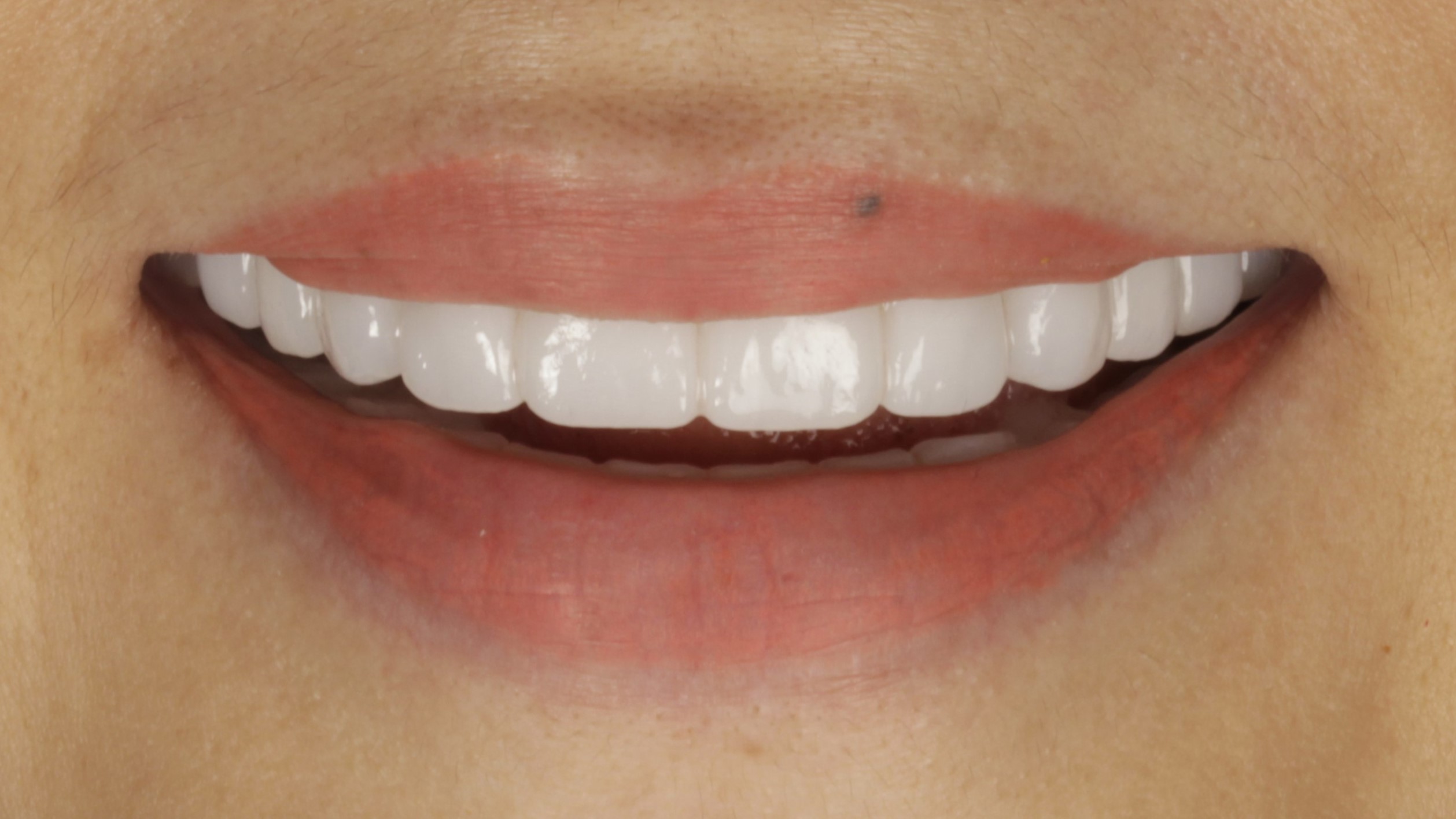 BeforeAfter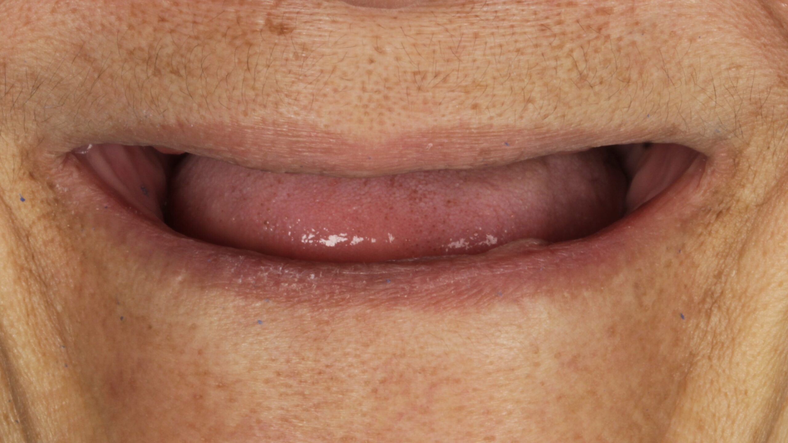
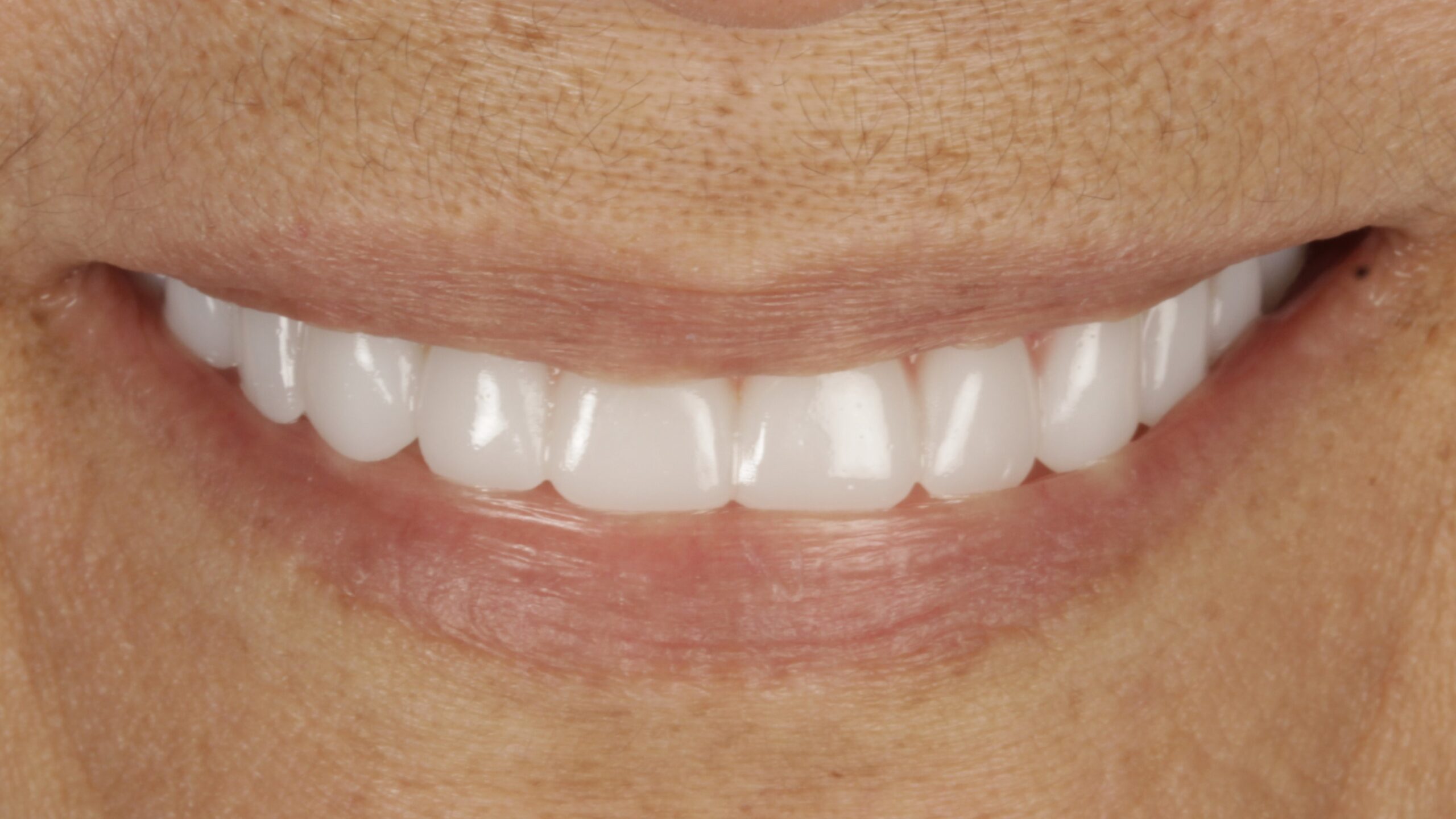 BeforeAfter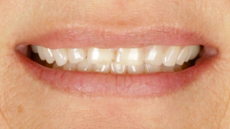
 BeforeAfter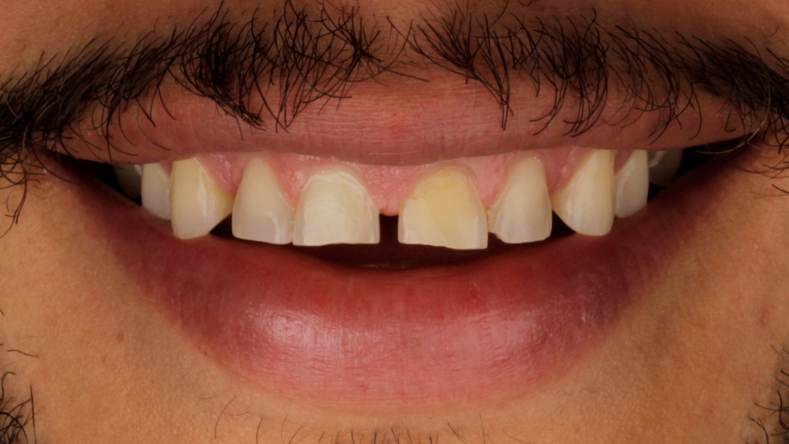
 BeforeAfter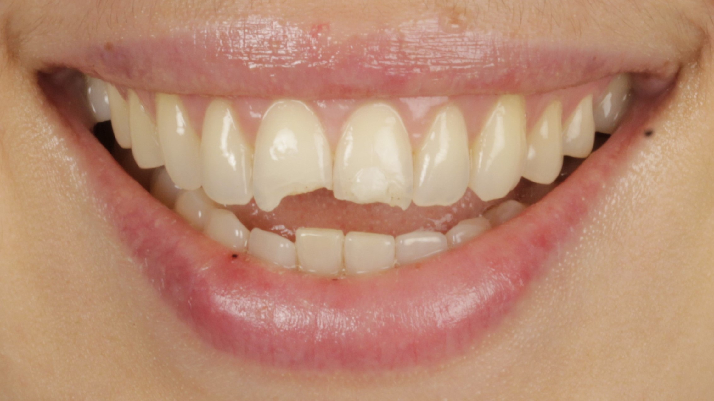
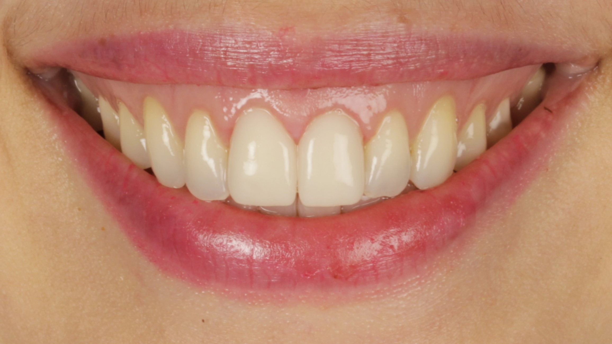 BeforeAfter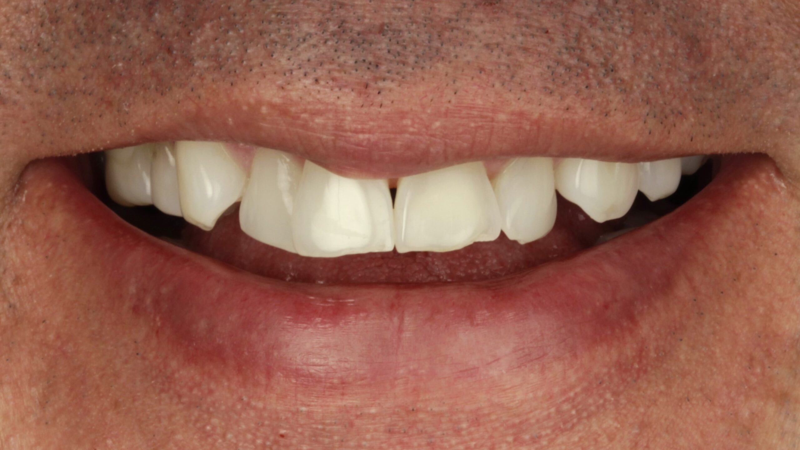
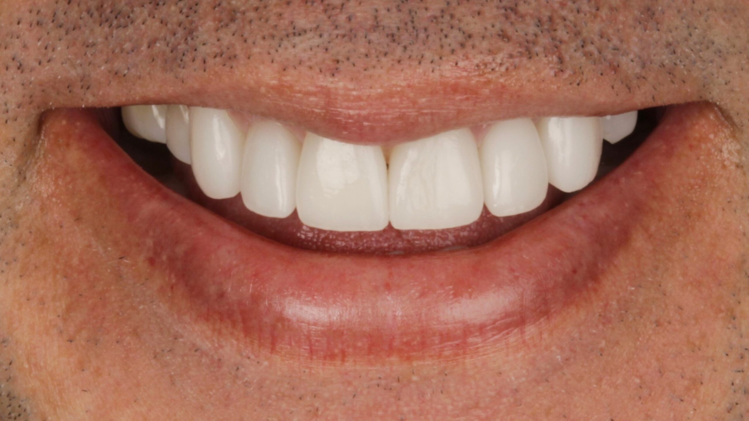 BeforeAfter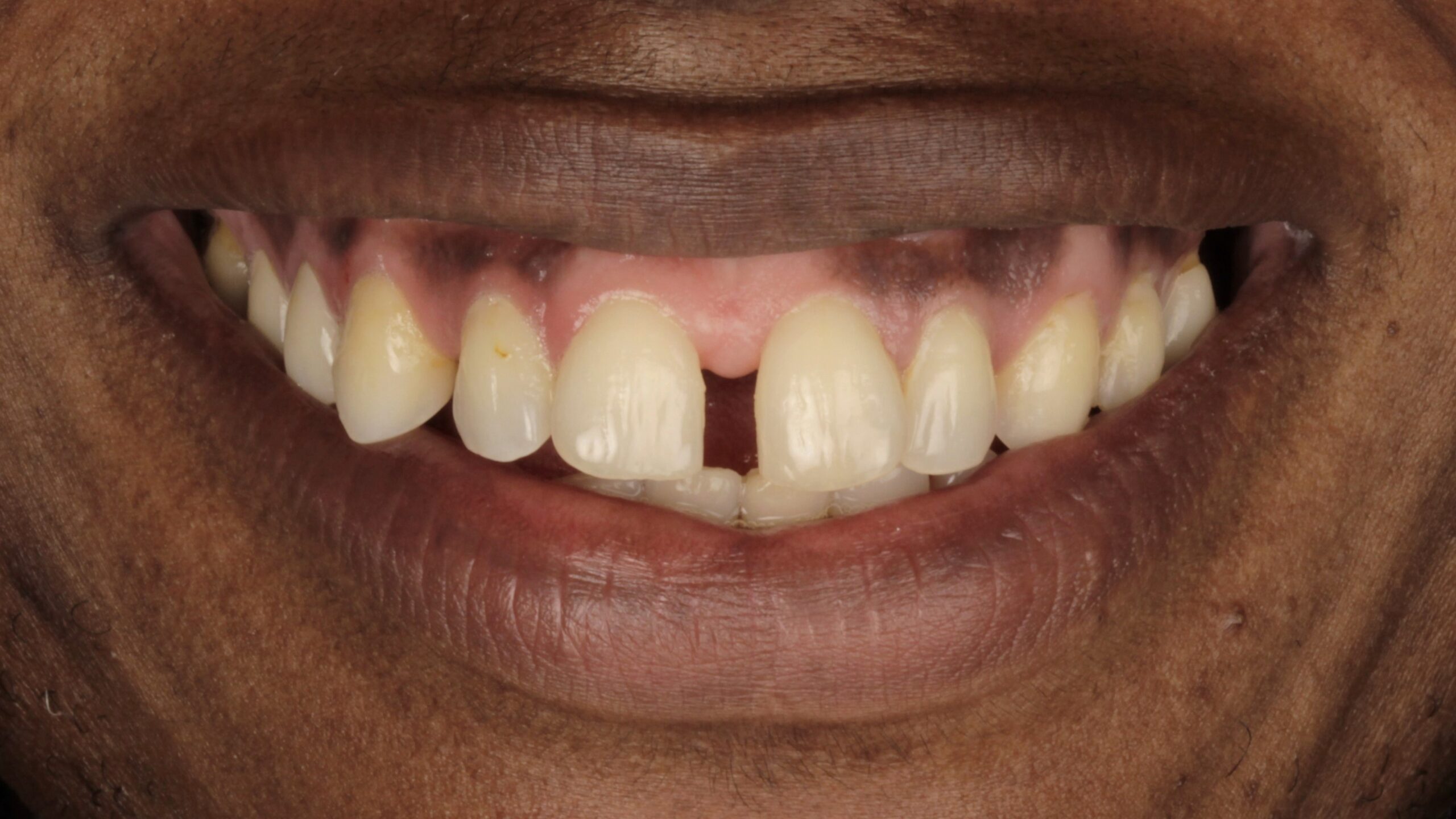
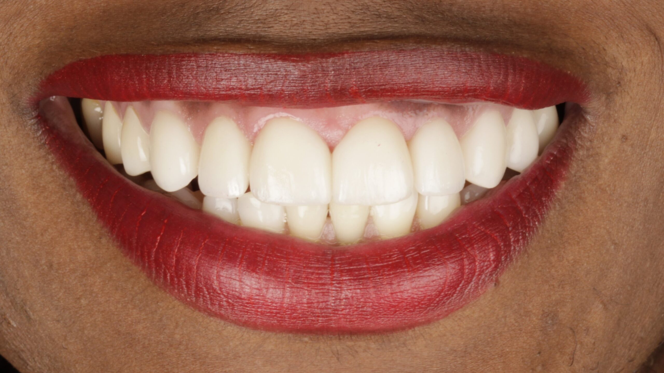 BeforeAfter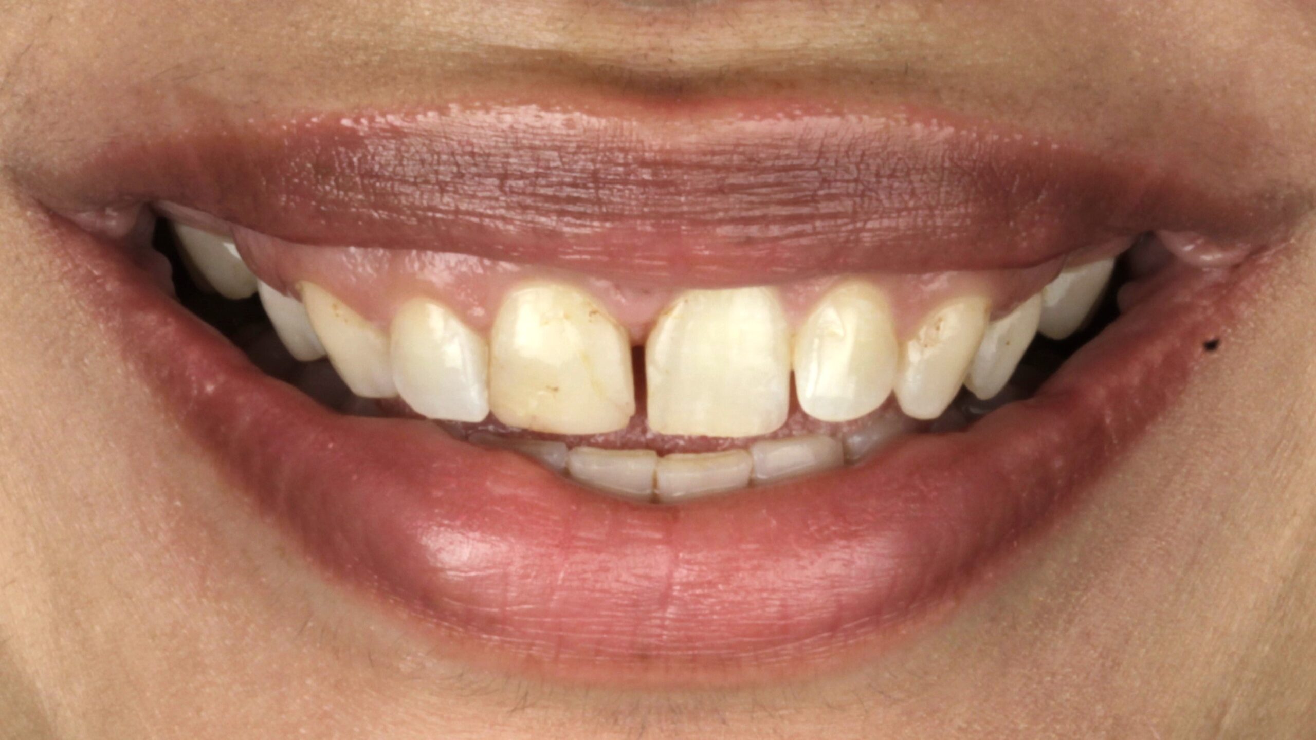
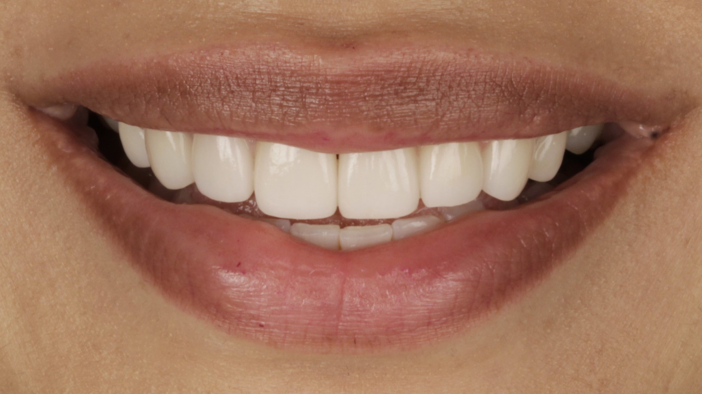 BeforeAfter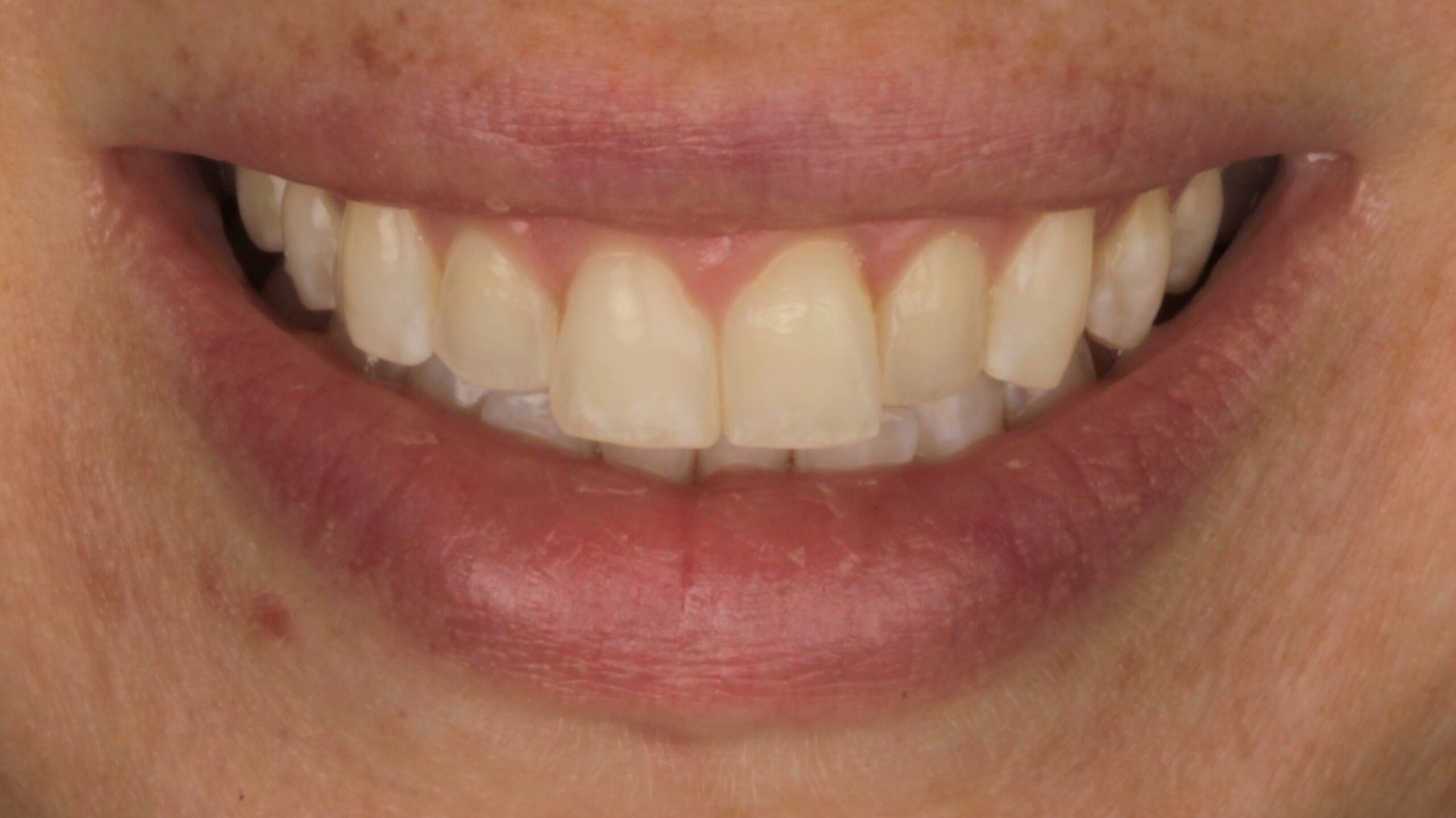
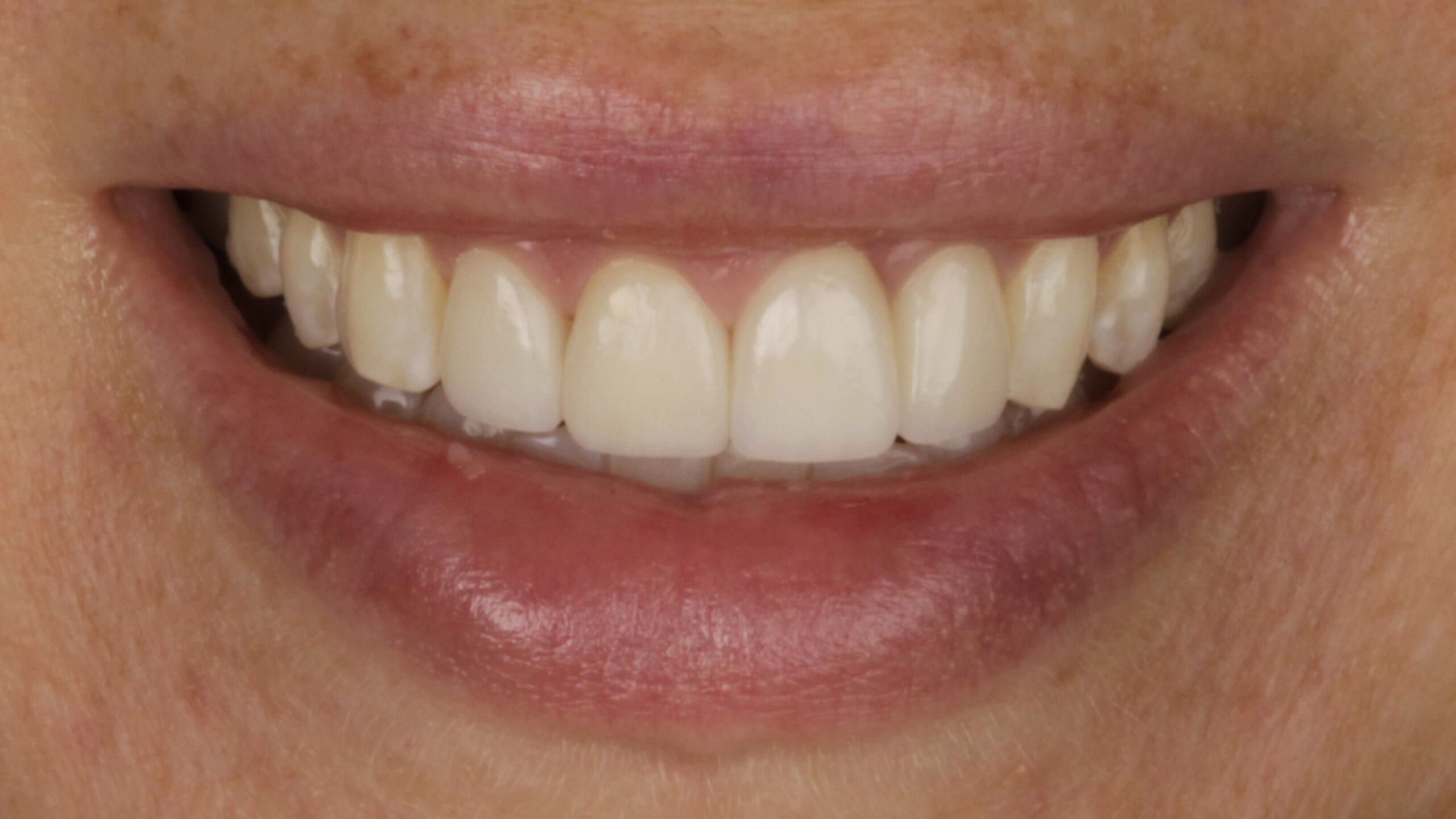 BeforeAfter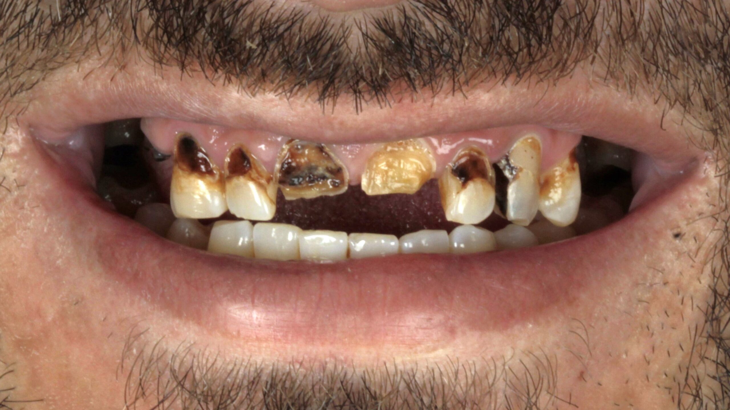
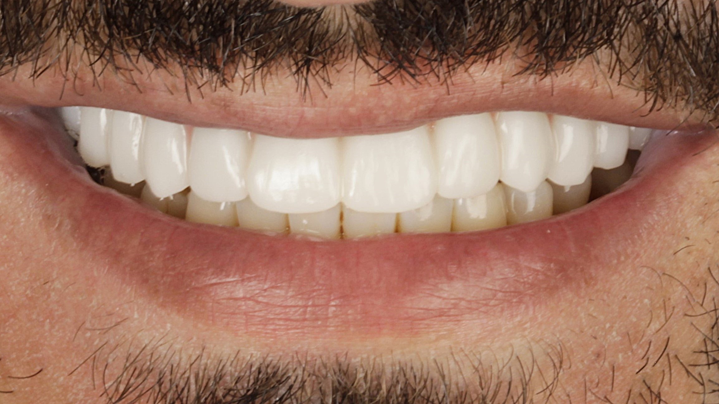 BeforeAfter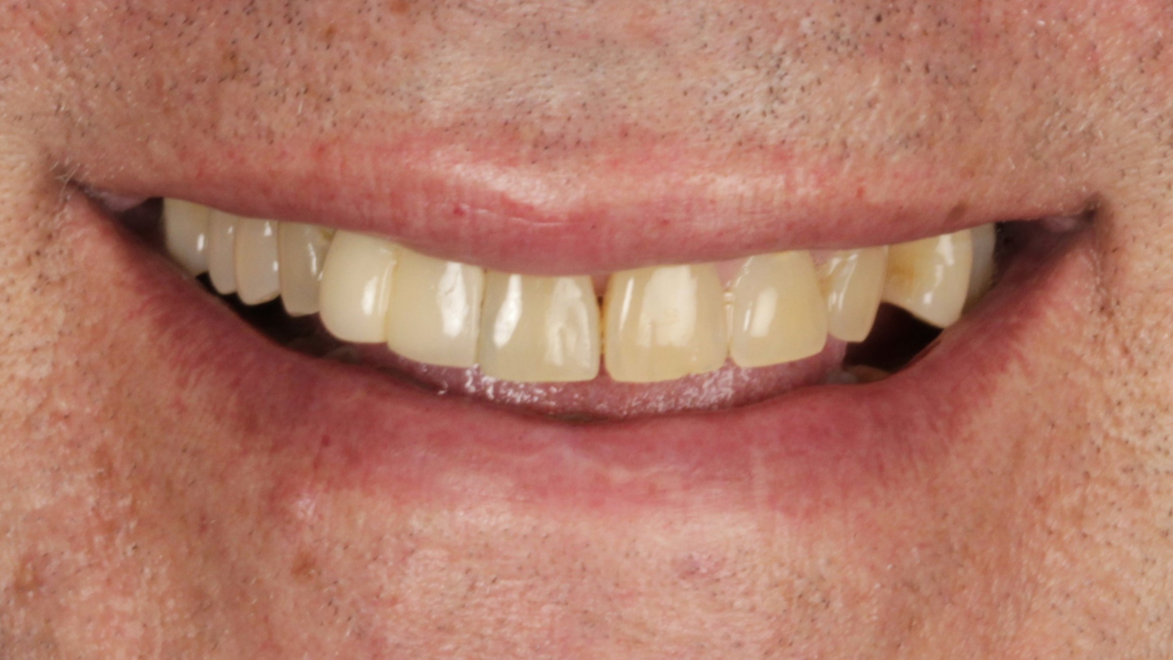
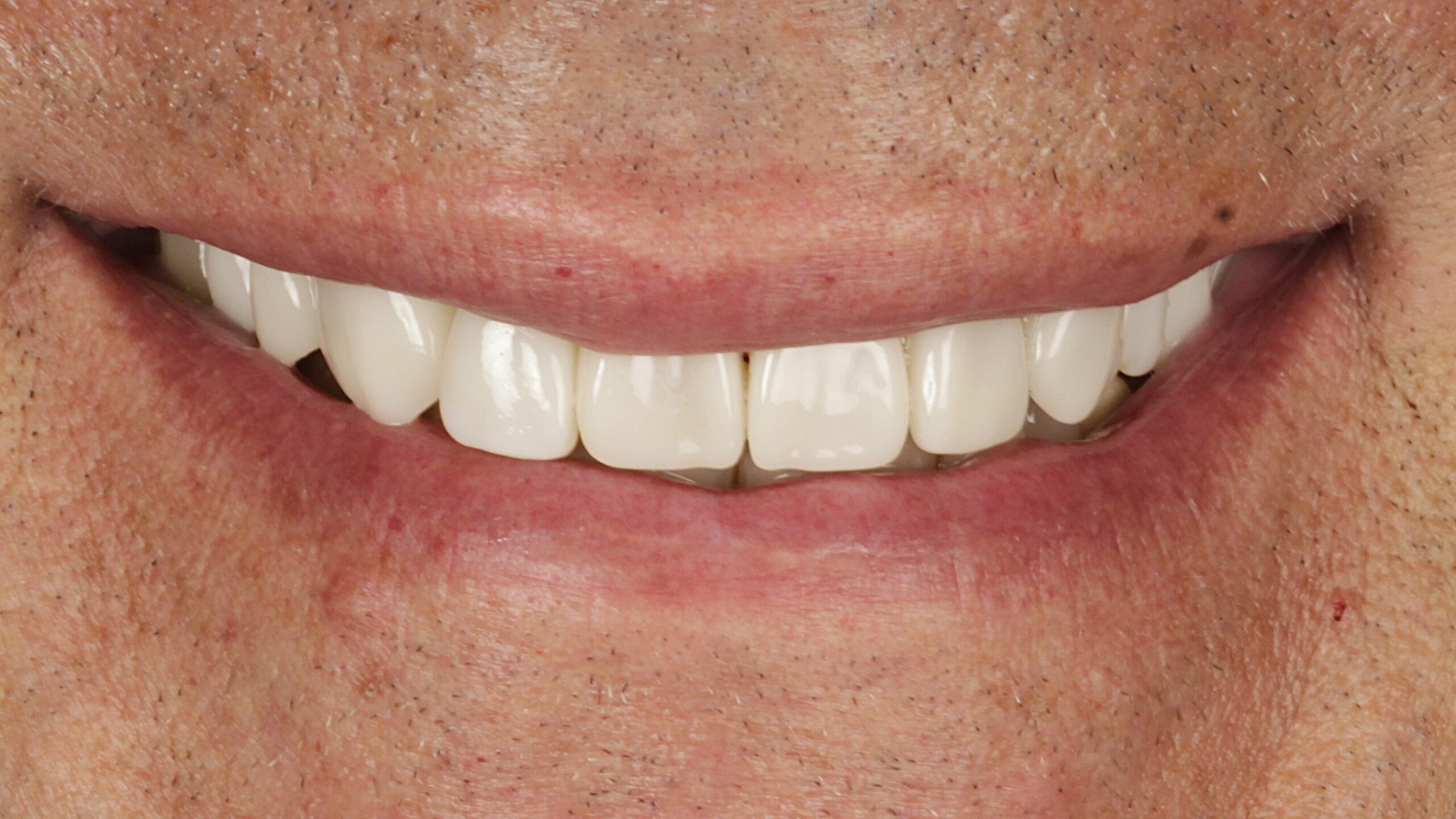 BeforeAfter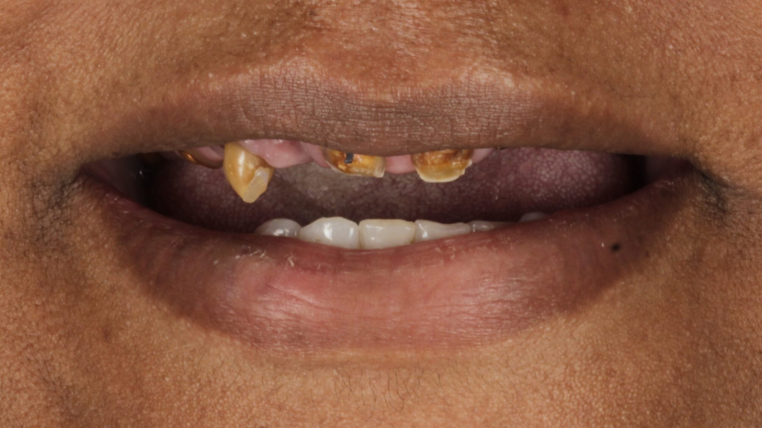
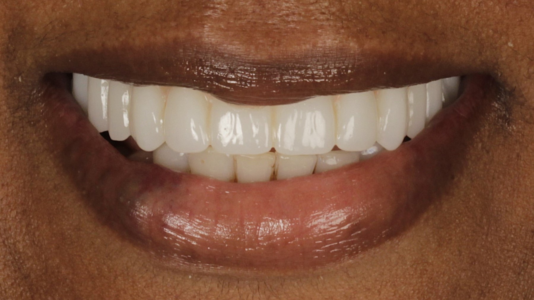 BeforeAfter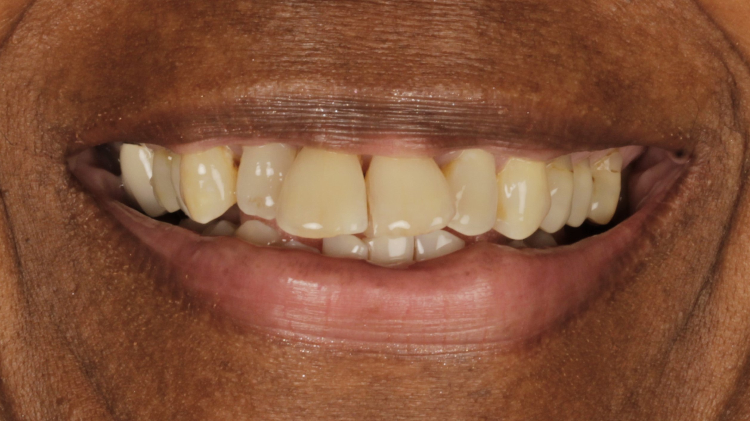
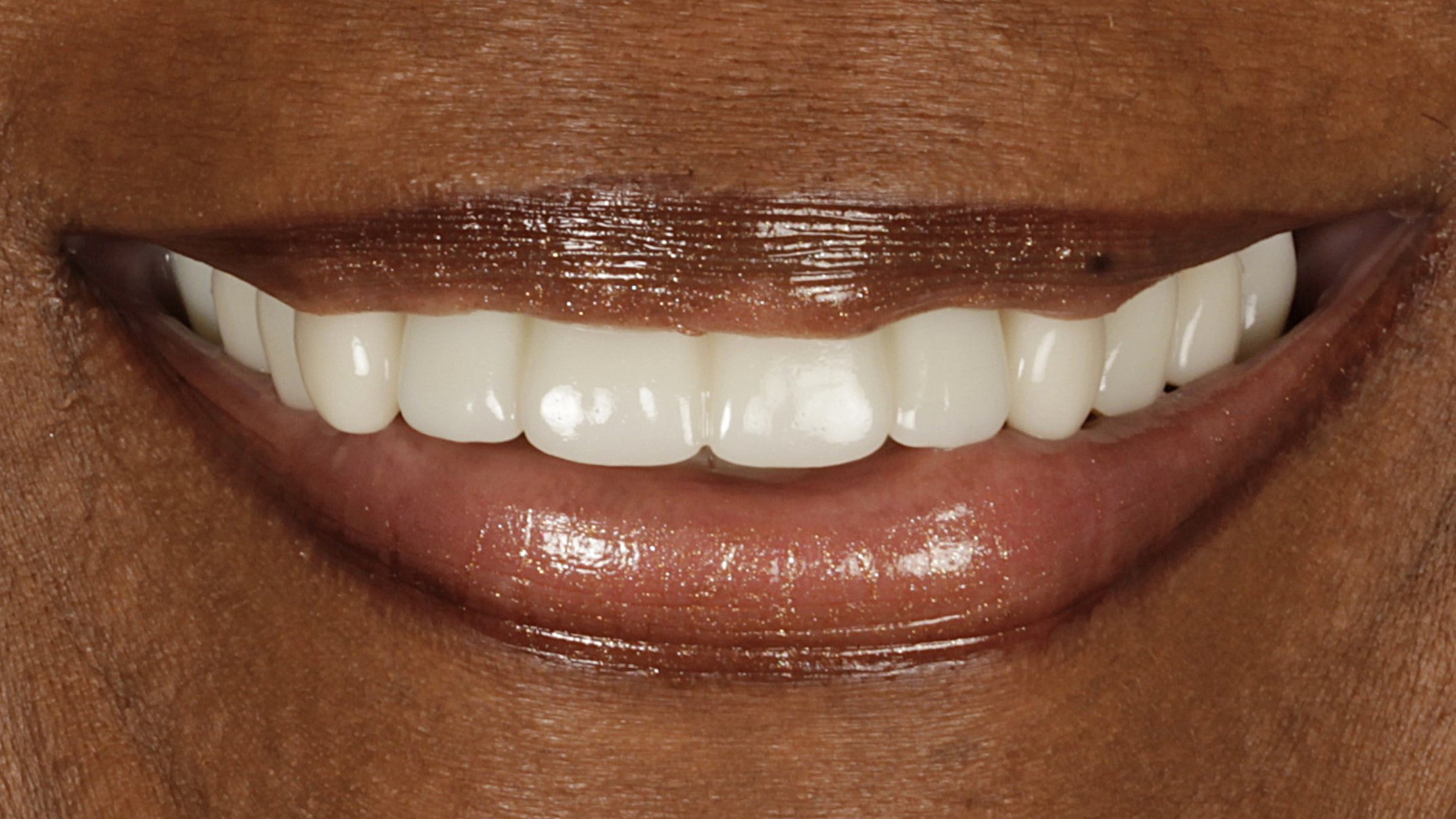 BeforeAfter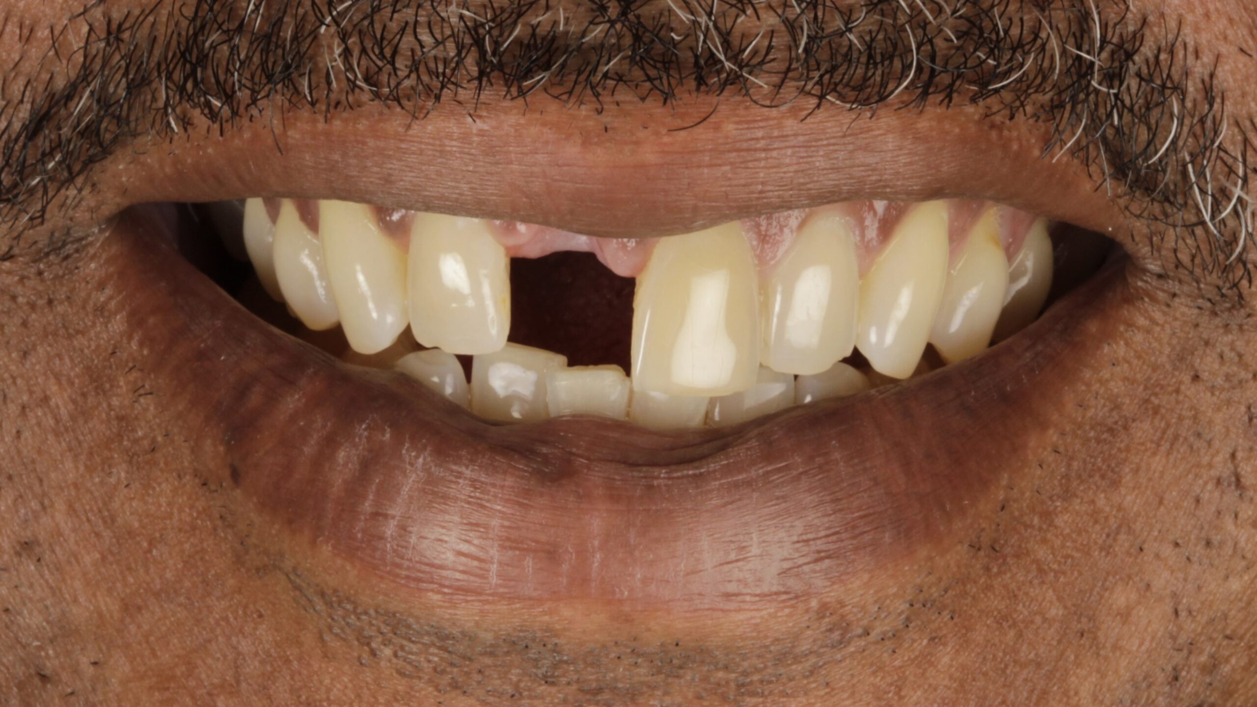
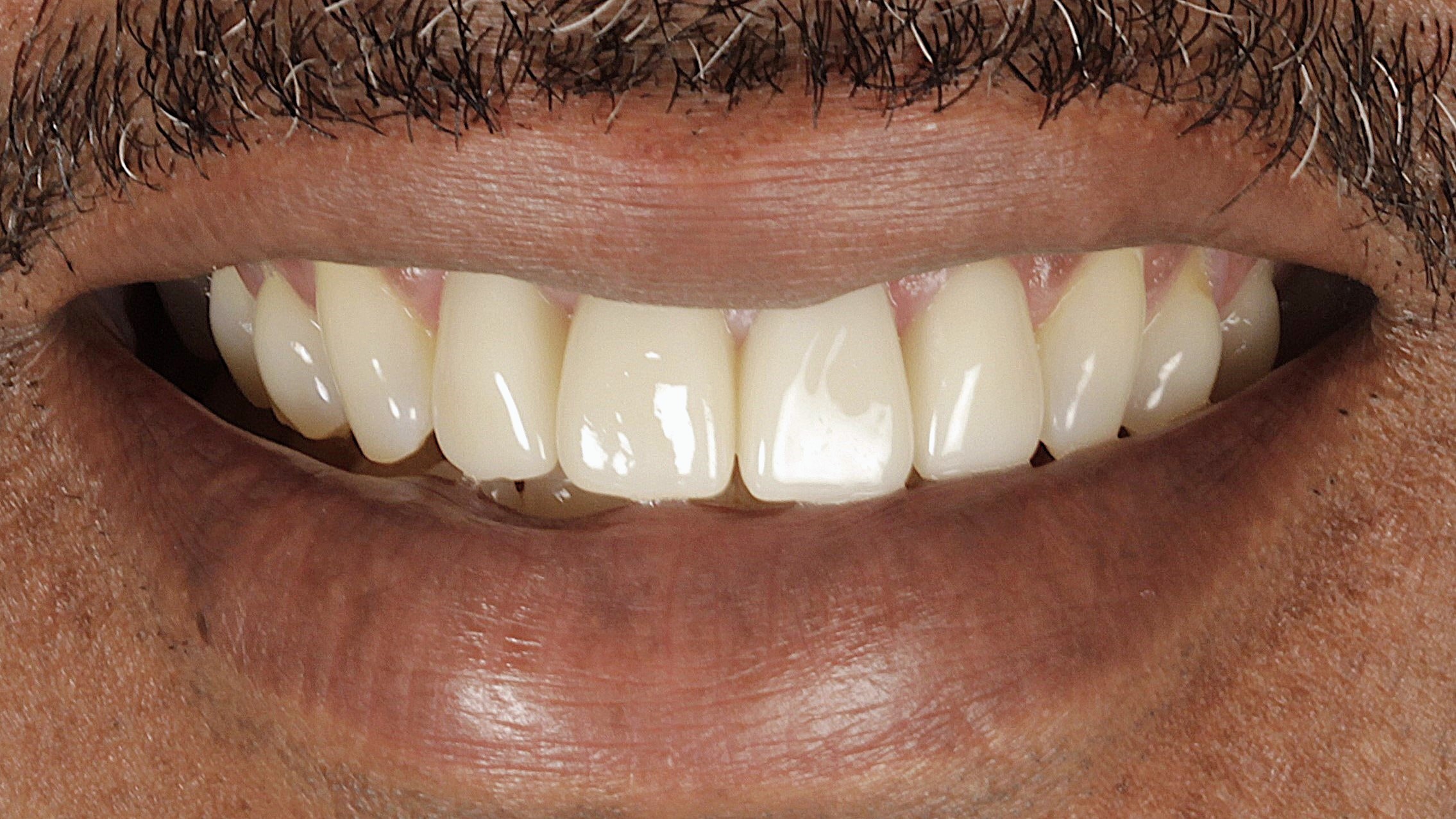 BeforeAfter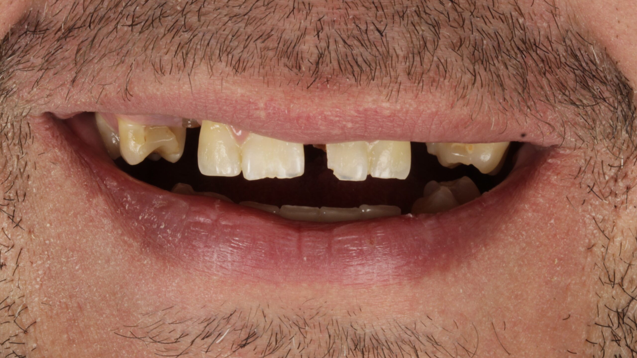
 BeforeAfter
 BeforeAfter
 BeforeAfter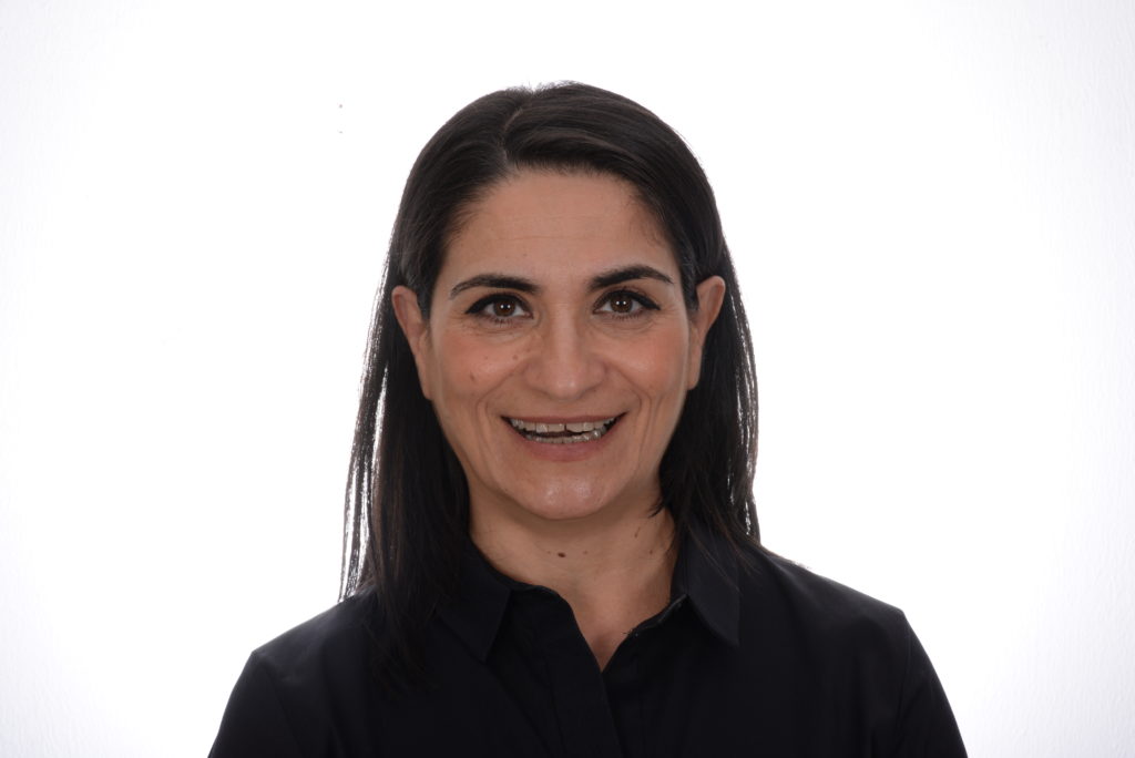
 BeforeAfter
 BeforeAfter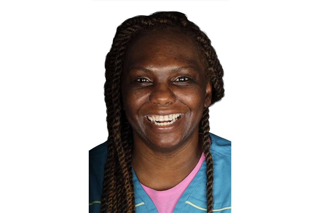
 BeforeAfter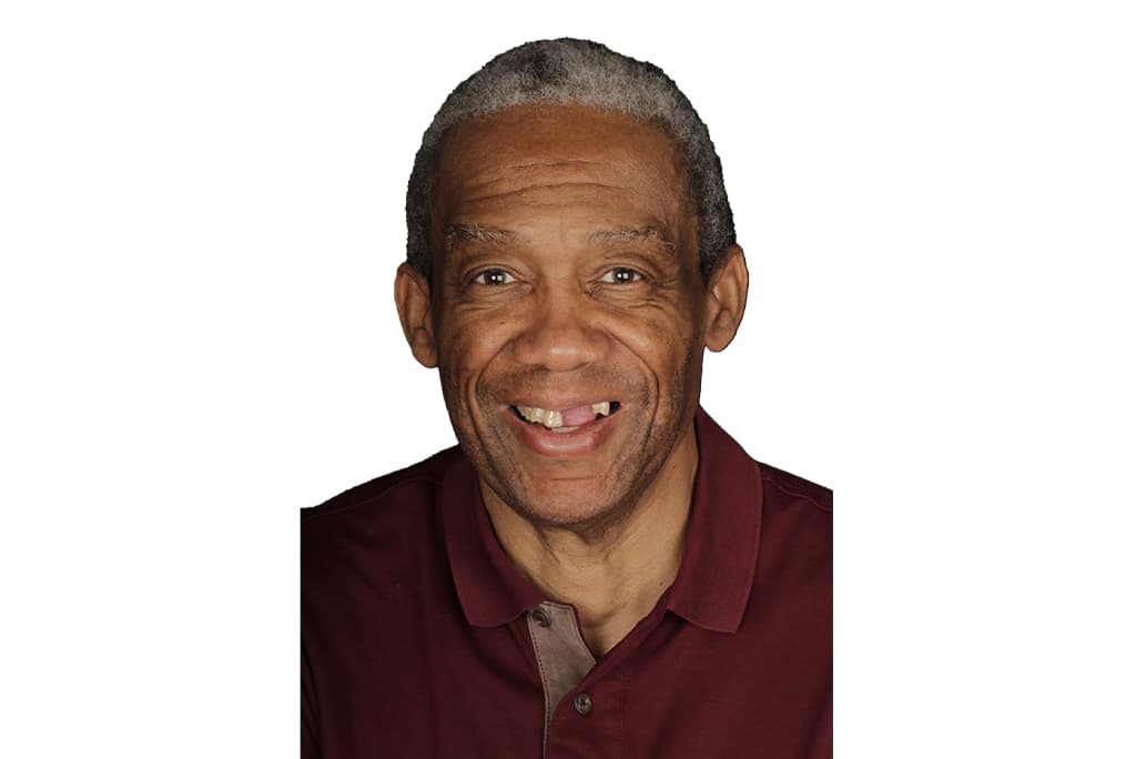
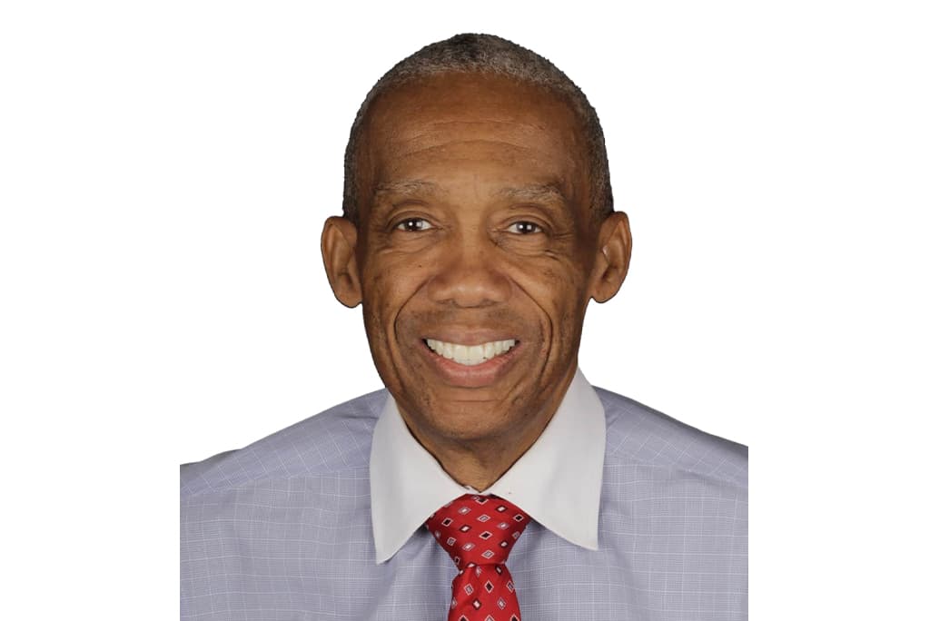 BeforeAfter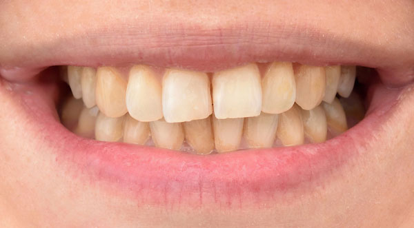
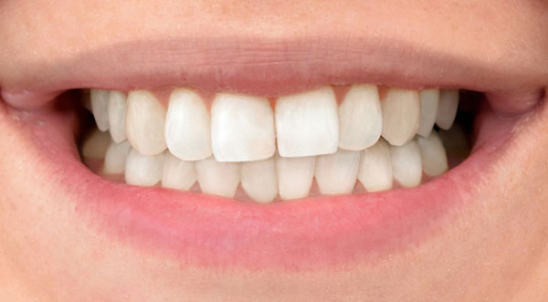 BeforeAfter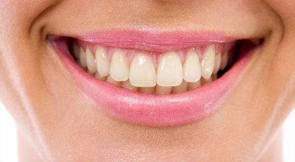
 BeforeAfter
What a before-and-after can and cannot tell you.
Clinical photographs are taken close, lit hard and shot at the same angle each time, which is why they look nothing like a portrait. That consistency is the point: it is what makes two images of the same mouth comparable.
What they cannot show is how the case was planned: how much bone there was to work with, whether teeth had to come out first, how the bite was balanced, how long it took. Two identical-looking afters can sit on completely different treatment plans.
So use these for what they are good for: seeing that work of this kind is done here, routinely, on cases other practices call too complex. Then bring your own mouth in and have it scanned.
Book a consultationWhere to go next.
Is your case in here?
Bring it in. The consultation includes a 3D scan and a straight answer about what your case actually needs.